Cell Biology

... from active transport to vesicles

CELL BIOLOGY

Cell Biology deals with the physiological function, structure, communication, reproduction, and death of cells. Cell Signaling relates to communication between cells. Molecular Biology deals with the ions and molecules involved in cellular functioning.

Overview of Cell Biology:

Prokaryotic cells of the Eubacteria and Archaea differ from eukaryotic cells in genetics, metabolism, and cell structure (lack of a nuclear membrane, lack of organelles, differences in cell walls). Prokaryotic flagellae and adhesion molecules participate in prokaryotic chemotaxis.

Table  Comparisons of Eubacteria, Archaea, and Eukaryotes  Cell walls of Prokaryotes  Electron acceptors for respiration and methanogenesis in prokaryotes  Glycolysis in bacteria  Lithotrophic prokaryotes  Comparison of Plant and Bacterial Photosynthesis  Structure of bacteriochlorophylls 

The cells of eukaryotic organisms are larger and structurally more complex than those of prokaryotes. Eukaryotic cells are divided into functional compartments (cytoplasm, organellar lumens, nucleus, vacuoles, vesicles) by a variety of cell membranes. Membrane-bound subcellular energy plastids (chloroplasts, mitochondria) were acquired through serial endosymbiotic events about 1 billion years ago. Cellular compartments include the nucleus, which communicates with the cytoplasm by way of nuclear pores and the endoplasmic reticulum. Cytoplasmic compartments include the cytoskeleton (microtubules, microfilaments, intermediate filaments, basal bodies and centrioles) specialized vesicular structures such as the endoplasmic reticulum, Golgi apparatus, proteasomes, lysosomes, plant vacuoles, endosomes and exosomes, and energy organelles (chloroplasts, mitochondria).

Cells perform a wide variety of physiological functions such as active transport and intracellular transport; proliferation, differentiation, the cellular stress response, and programmed cell death; they respond to the environment through chemotaxis, energy transduction, and signaling; and move materials into the cell through phagocytosis, pinocytosis, and receptor-mediated endocytosis.

Cell membranes comprise phospholipids, sugars, and specialized proteins and perform a variety of functions such as adhesion, cellular communication, and maintenance of concentration gradients by active transport. These activities procede by means of specialized adhesion molecules, signaling molecules, ion channels and pumps, and receptor proteins.

The cell's cytoskeleton participates in chemotaxis, intracellular transport, and cell-to-cell adhesion. The cytoskeleton interacts with the extracellular environnment by way of transmembrane adhesion molecules (CAMs) and through surface protuberances such as cilia and flagellae (with various functions as mechanoreceptors, chemoreceptors, and the outer segment of the rods in the vertebrate retina). The centriolar components of the cytoskeleton organize the spindle apparatus on which the nuclear chromosomes translocate during mitosis and meiosis, phases of the reproductive cell cycle.

In multicellular organism, proliferation of cells must be balanced by cell death, usually through programmed, non-inflammatory apoptosis.

 Cell Adhesion Molecules  Immune Cytokines  Second Messengers  Cell signaling
 Apoptosis vs Necrosis  Apoptosis 

This site deals with Cell Biology, Cell Signaling, and the Molecular Biology of cells, particularly in relation to genetic mechanisms of biological evolution. Blue terms hyperlink to explanatory items so that you may navigate through items providing more detail. The site is searchable using the "Search this blog" box at top left. Use the "back" function to return to each departure item.

Alphabetic listing of Items - main items are bold
CELL BIOLOGY : A : active transport : adhesion : apoptosis : C : cell cycle : cell membranes : centrioles : (chemical gradients) : cilia and flagella : communication : concentration gradients : cytoplasm : cytoskeleton : D : death of cells : E : endocytosis : endoplasmic reticulum : endosomes : energy transducers : eukaryotic : exosome : F : flagella : G : Golgi apparatus : I : ion channels : L : lysosome : M : meiosis : microtubules : mitochondrion : mitosis : mitotic spindle : N : nuclear membrane : nuclear pore : nucleolus : nucleus : P : peroxisome : transport : phagocytosis : photosynthesis : physiological function : pinocytosis : plant cell : plasma membrane : prokaryotic : proteasome : protein degradation : protein pumps pumps : R : receptor proteins : receptor-mediated endocytosis : reproduction : ribosomes : RTKs : S : spindle : structure : V : vacuole : vesicle :

CELL SIGNALING : AKAPs : cellular signal transduction : concentration gradients : chemical gradients & communication : chemotaxis : cytokines : cytokine receptors : DAG : DAGKs : diacylglycerol : diacyl glycerol kinase : ERKs : GPCRs : GPCR families : hormones : neural action neural activity neuronal activity neuronal interconnections : neurotransmission : neurotransmitters : Nitric Oxide : PDZ domain : phosphorylation : phosphotransfer-mediated signaling pathways : PKA, protein kinase A : phospholipase C-gamma : PLC-G : PKC : protein kinase A : protein kinase C : protein tyrosine kinases protein kinases : Protein Kinase Signaling Networks : Ras : second messengers : signaling gradients (concentration gradients : chemical gradients) : signal transduction : two-component systems :

MOLECULAR BIOLOGY : amino acids : caspases : (concentration gradients) : (hormones) : proteins : Chemistry of Life site Biochemistry & Molecular Genetics : Molecular Genetics site

Items occur within Sections (listed in the sidebar). Items are listed in groups of 10 – to see more items, click on the lowest item in the sidebar. When visiting an item, the site title changes to purple – click on the title or “Home” to return to the main page.

The “Guide-Glossary” link below each item provides a glossary of terms ( # > 0 ), as a pop-up when reading within a Section, or as sub-script when visiting an Item.

| 0 Guide-Glossary

Inner Life of the Cell

For any who have not yet realized that the biology of cells can be fascinating – and aesthetic – Harvard University has sponsored an animation of the "Inner Life of the Cell" (HiRes, LoRes).


Unfortunately, the Internet-released version lacks a commentary soundtrack, settling instead for a music background that restricts the experience to pure aesthetics in the absence of explanation. My attempt at interpretation follows:

The animation opens with the rolling adhesion of a leukocyte within a blood vessel. The surfaces of two cells are shown adhering at contact points between adhesion molecules (selectin-saccharide).

We enter the cell and see a lipid raft, with its embedded sphingolipids and phosphatidyl choline(cholesterol+proteins), floating within the plasma membrane. Next we see a multi-protein focal contact, and then the cytoskeleton. After glancing back at the sub-plasma membrane 'geodesic' microfilaments, we pass down throught the cytoskeletal lattice and see actin microfilaments assembling, then depolymerizing after attachment of a protein (gelsolin?).

Next we see assembly and disassembly of tubulin. This interesting sequence is followed by my favorite segment, which is kinesin dragging an endosomal vesicle along a microtubule as kinesin-bigfoot 'walks' along the tubulin that is radiating from a centriole pair. A fellow kinesin, in the background, actively transports an endosome in the opposite direction.

Next, we approach the nuclear envelope with its embedded nuclear pore complexes. Several mRNA molecules with attached proteins exit the nucleus through the pores and assemble into loops within the cytoplasm, where the mRNA is scanned for a start codon, and is then translated into new polypeptide/protein chains by a ribosome. Globular proteins dimerize and tumble toward a mitochondrion.

Further translation injects a nascent protein chain through a pore into the endoplasmic reticulum as 'bigfoot' continues to clomp along tubulin, dragging a vesicle behind. Next, we see the Golgi apparatus budding vesicles before our plodding kinesin reaches the plasma membrane and proteins are released into the ECF by exocytosis. The newly synthesized integrins float on a lipid raft before unfolding into their active conformation, whereupon they snare a passing leukocyte and induce extravasation.

Happily, an extended version of the animation, complete with commentary, is now available on YouTube.


See a video of medical animator David Bolinsky describing the making of Inner Life of the Cell. More Cell Biology Videos.

The creationist mucusballs at the Dissembly Institute have plagiarized the footage, renamed it The Cell as an Automated City (presumably because that analogy is within the conceptual grasp of creationists), removed its credits, and are passing it off (implied) as Behe's research into the complexity 'created' by the IDer (aka God). The IDiots collect donations from credulous creationists as the DI Fellows pretend to conduct scientific investigation of IDiocy. I sincerely hope that Harvard University and XVIVO sue the pants of the IDiots, just as I'd like to see them tried and convicted of their plagiarism at the Kitzmiller vs Dover School Board trial.
See Creationist crooks pilfer Harvard's work . DI Fellows-- EXPELLED for plagiarism . Plagiarism and Intelligent Design .

Labels: , , , , , , , , , , , , , ,

| 0 Guide-Glossary

Fantastic Voyage

The following talk Fantastic voyage inside a cell was presented by medical animator Ted Bolinsky at TED, who describes the creation of Inner Life of the Cell. 9 min 57 secs.


Labels: , , , , ,

| 0 Guide-Glossary

Cell Biology Videos

On Cell Biology:
Inner Life of the Cell : Fantastic Voyage :

On Chemistry of Life:
transcription :

On YouTube:
The Inner Life of the Cell : Inner Life of a Cell: Leukocyte : Harvard Biovisions - The Inner Life of a Cell : Nano Visualization / Micro reality - Life Inside a Cell : Beginning of Life : CELL wrapping & DNA replication : CELL wrapping & DNA replication : THE MIRACLE OF MAN'S CREATION : Cell Signals (Part 2 of 2) : Sample clip from "Voyage Inside the Cell" V035 : The Life...of a cell. :

Labels: , , , , ,

| 0 Guide-Glossary

active transport

Active transport pumps require expenditure of energy, most often in the form of ATP, to transport hydrophilic macromolecules and ions across membranes against chemical or concentration gradients. The number of integral protein transporters in the membrane limits active transport.

Primary, or direct transport involves an energy expending conformational change in the membrane protein to transport a specific molecule or ion across the membrane. Secondary, or indirect transport utilizes energy directly to generate a transmembrane gradient down which ions move and then up or down which the coupled molecule or ion of interest is transported indirectly.

ATPases couple the hydrolysis of ATP (to ADP and Pi) with the transmembrane transport of ions against a concentration gradient. An example is Na+K+ ATPase, which pumps 3 Na+ ions out of the cell for 2 K+ ions it pumps into the cell. Because the pump moves ejects three Na+ for every two K+ moved inward, it generates a net electrical differential necessary for polarization. This electrical potential energy is essential for neuronal activity, and it supplies the energy needed for other types of transport such as symport and antiport. animation - Na K ATPase

Active transport of some substances against concentration gradients employs the ATPase-derived energy stored in ion gradients, such as proton (H+) or sodium (Na+) gradients, to drive transporter membrane proteins. In a symport, the ATPase transported molecule and the coupled, co-transported ion move in the same direction. Conversely, the ATPase transported molecule and the coupled, co-transported ion move in the opposite direction in an antiport.

Translocases utilize energy to move large molecules across the outer (Tom complex) and inner (Tim complex) mitochondrial membranes [image, more detail].

Џ beautiful Flash 8 animation - Inner Life of the Cell, which shows active transport of endosomes, and Interpretation: Inner Life of the Cell

Harvard University provides a wonderful video explaining operation of the F1-F0 ATPase. Џ

• A • adhesion • C • cell membranescellular signal transductioncentrioleschemotaxischloroplastciliacommunicationconcentration gradientscytokine receptorscytoplasmcytoskeleton • E • energy transducersendoplasmic reticulumendosomesexosome • G • Golgi apparatusGPCRs • H • hormones • I • ion channels • L • lysosome • M • meiosismicrotubulesmitosismitochondrion • N • Nitric Oxideneurotransmissionneuronal interconnectionsnuclear membranenuclear pore • P • pinocytosisproteasomepumps • R •receptor proteinsreceptor-mediated endocytosis • S • second messengerssignaling gradientssignal transductionspindlestructure • T • transporttwo-component systems • V • vacuolevesicle

animation - Na glucose symport : Carriers, Pumps, & Channels : The Virtual Cell Textbook - Cell Biology

Labels: , , , , , , , , ,

| 0 Guide-Glossary

adhesion

The cells of most eukaryotic species adhere to one another in co-operative multicellular organisms, whereas prokaryotes are unicellular organisms even though some species form colonies. Cell adhesion relies upon specialized transmembrane cellular adhesion molecules (CAMs) that usually extend from the intracellular space to the extracellular space where they may bind to other cell membranes or to the extracellular matrix. Within the intracellular domain, adhesion proteins adhere directly to, or are coupled to, the cell's cytoskeleton. Cadherin (calcium dependent adhesion) molecules are normally coupled by special linking proteins – catenins – to the cytoskeleton.

Intermediate filaments are about 10 nm diameter and provide tensile strength for the cell and connect adjacent cells through desmosomes (macula adherens).
cytoskeleton : diagram . desmosome : tem_desmosome : diagram . tight junction : diagram . gap junction : image - cytoskeleton : image_cytoskeleton : diagram - mechanism of ciliary motility Џ beautiful Flash 8 animation - Inner Life of the Cell, which shows the sequelae of adhesion-signaling, and Interpretation: Inner Life of the Cell Џ

 Cell Adhesion Molecules  Second Messengers  Cell signaling

There are several families of adhesion proteins, each with specific ligands of the same type (homophilic) or a different type (heterophilic).
1. integrins with heterophilic attachments to different (hetero) ligands in the extracellular matrix
2. selectins with heterophilic attachments to carbohydrate ligands
3. Ig superfamily proteins with selectins with heterophilic attachments to: a) integrin ligands, and b) Ig superfamily proteins of a different (hetero) type, and
homophilic attachments to the Ig superfamily proteins of the same (homo) type
4. cadherins with homophilic attachments to cadherins of the same type, or by way of catenins (right - click to enlarge, description), to the cytoskeleton

Cell adhesion is important in:
1. maintaining contact within solid tissue
2. embryogenesis (morphogenesis)
3. migration of single cells such as leukocytes within multicellular organisms
4. maintaining contact between neuronal elements
5. virulence of virions and bacteria

Some signaling molecules act as adhesion receptors, and cluster in focal adhesions upon ligand binding. (Rho protein). A variety of integrins, which are transmembrane heterodimeric adhesion receptors are known to support adhesion-dependent growth factor-activation of MAP kinase. Focal adhesions are rich in tyrosine phosphorylated proteins, coupling cell adhesion to signal transduction pathways in the cell. Various adhesion receptors, such as integrin, are closely linked to protein kinases and phosphatases. Grb2 links focal adhesion kinase (FAK) to the Ras pathway when Grb2 is phosphorylated after binding to FAK. The 85 kDa subunit of the PI 3-kinase is also phosphorylated after binding to FAK. Thus, FAK is a key component in the assembly of focal contact structures that influence cytoskeletal organization and signal transduction.

Engagement of ICAM-1, a member of the immunoglobulin supergene family (Ig), has been documented to activate specific kinases through phosphorylation, resulting in activation of transcription factors, increased cytokine production, increased cell membrane protein expression, production of reactive oxygen species, and cell proliferation.

cadherins : Ig superfamily proteins : integrins : selectins : ICAMs :  Cell Adhesion Molecules
 Second Messengers  Cell signaling .

Cells form a variety of intermembranous junctions: adherens junctions, desmosomes (plasmodesmata), focal contacts, gap junctions, hemidesmosomes, tight junctions.

cytoskeleton : diagram . desmosome : tem_desmosome : diagram . tight junction : diagram . gap junction : image - cytoskeleton : image_cytoskeleton : diagram - mechanism of ciliary motility :

• A • adhesion • C • cell membranescellular adhesion moleculescellular signal transductioncentrioleschemotaxischloroplastcilia & flagellacommunicationconcentration gradientscytokine receptorscytoplasmcytoskeleton • E • energy transducersendoplasmic reticulumendosomesexosome • F • flagella & cilia • G • Golgi apparatusGPCRs • H • hormones • I • ion channels • L • lysosome • M • meiosismicrotubulesmitosismitochondrion • N • Nitric Oxideneurotransmissionneuronal interconnectionsnuclear membranenuclear pore • P • pinocytosisproteasomepumps • R • receptor proteinsreceptor-mediated endocytosis • S • second messengerssignaling gradientssignal transductionspindlestructure • T • transporttwo-component systems • V • vacuolevesicle

| 0 Guide-Glossary

apoptosis

Cellular death-by-suicide is part of normal development, and is termed apoptosis or programmed cell death (PCD). Cysteine Aspartate Specific ProteASEs – caspases – are active in apoptosis, as are p53, a tumor suppressor gene, and FAS gene, which is member 6 of the tumor necrosis factor receptor superfamily (TNF). In contrast to apoptosis, necrosis is cell death that results from cytotoxic, injurious stresses that are too severe for correction by the cellular stress response.

▼: AIF : ANT : apoptotic appearance : apoptosomes : apoptotic bodies : Bcl-2 : Bcl-2 family : caspase cascadecaspases : cytochrome C : death domain : death receptor pathway : DISC : Endonuclease G : FADD : FAS gene : homeostasis and apoptosis : MBR : mitochondrial pathway : necrosis : PBR : porin : proteolytic cascade : PT pore : TNF-R : tumor suppressors : Smac/DIABLO : VDAC : ▼

Tumor suppressor proteins such as p53 act as cell-cycle repressors and/or promoters of apoptosis through:
1. interruption of cell cycle, preventing cell division,
2. halting the cell cycle if DNA damage is not yet repaired,
3. inducing apoptosis if DNA damage cannot be repaired,
4. promoting cell adhesion and contact inhibition, which prevent invasion and metastasis.

Both the death receptor pathway and the mitochondrial pathway lead to the activation of an initiator caspase, which initiates a proteolytic cascade, ultimately leading to apoptotic cell death.

Necrosis results from impairment of the cell’s ability to maintain homeostasis due to breakdown of the plasma membrane. This leads to influx of water as pumps fail and osmotic gradients reverse. Ultimately organelles (especially mitochondria) swell and hydrolytic, lysosomal enzymes are released into the cytosol, leading to cellular swelling and rupture (lysis). So, in vivo, necrotic cell death is often associated with extensive tissue damage resulting in an intense inflammatory response.

Apoptosis, by contrast, occurs under normal physiological conditions when the cells respond to signals as active participant in their own deaths. Apoptosis is a part of normal cell turnover and tissue homeostasis. It plays a significant role in embryogenesis, induction and maintenance of immune tolerance, development of the nervous system, and atrophy of endocrine-dependent tissue.

[] Artist's impression apoptosis []

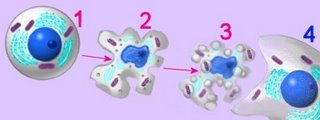
Cells undergoing apoptosis show characteristic morphological and biochemical features, which include chromosomal changes: chromatin aggregation, nuclear and cytoplasmic condensation (2), and partition of cytoplasm and nucleus into apoptotic bodies (apoptosomes). These are membrane bound-vesicles that contain ribosomes and intact mitochondria and nuclear material (3). Apoptotic bodies are rapidly recognized in vivo and and are phagocytozed by either macrophages or adjacent epithelial cells (4), without inflammatory response. In vitro, apoptotic bodies and remaining cell fragments ultimately swell and lyse (“secondary necrosis"). [more] Џ apoptosis animations Џ

Tables  Apoptosis vs Necrosis  Apoptosis 

Death receptor pathway:
Death receptors include the TNF-R (tumour necrosis factor receptors) and CD95 (Apo-1 or Fas) receptor families. Ligands for these death receptors are termed TNF-α and CD-95L (FasL), respectively. TNF-α is a highly cytotoxic molecule and the TNF-R1 receptor is widespread, with the result that TNFα has a low tolerated dose. Several members of the TNF-R1 family share a homologous region known as the death domain (DD), which is a protein-protein interaction domain that binds to adaptor proteins such as the adaptor protein FADD (Fas-Associated Death Domain). The Death-Inducing Signalling Complex (DISC) comprises death receptor ligands, death receptors and adaptor proteins such as FADD. Activation of death receptors by binding of ligands such as CD-95L (FasL) and TNF-α leads to caspase-8 activity.

Mitochondrial pathway:
Mitochondria play a central role in apoptosis and display an increase in mitochondrial membrane permeability during apoptosis. Mitochondria participate in apoptosis, and may be necessary for induction of apoptosis by apoptotic stimuli such as DNA damage. Pro-apoptotic and anti-apoptotic members of the Bcl-2 family are believed to regulate the release, through the mitochondrial PT pore, of pro-apoptotic substances such as AIF, Endonuclease G, Smac/DIABLO and cytochrome C. Mitochondrial efflux of cytochrome-c drives generation of the apoptosome (apoptotic body) in the cytoplasm. This in turn leads to caspase-9 activity.

Bcl-2 family: Members of the Bcl-2 subfamily (Bcl-2, Bcl-xL, Bcl-w) are localized on the outer mitochondrial membrane and show anti-apoptotic activity. They possess all four BH (Bcl-2 Homology) domains (BH1 to BH4). (Anti-apoptotic members of the Bcl-2 family have all four BH domains, while pro-apoptotic members have less BH domains.) Bax subfamily (Bax, Bak, BAD) : Bax is Bcl-2 Associated X protein, a pro-apoptotic member of the Bcl-2 family, which contains BH1 to BH3. BH3 only subfamily: Bid or tBid is (truncated) BH3-Interacting Domain death agonist, is a soluble, pro-apoptotic member of the Bcl-2 family, which contains only the BH3 domain.

Pro-apoptotic proteins : Bad, Bax, Bid, Bak, Bik, Bim, Bmf, Bok, Puma.
Anti-apoptotic proteins : Bcl-2, Bcl-XL, Mcl-1, Bcl-w

Released through the Mitochondrial Permeability Transition Pore (PT pore) are:
1. Apoptosis Inducing Factor (AIF) is a flavoprotein that is translocated to the nucleus where it causes DNA fragmentation into fragments of about 50kb.
2. Endonuclease G (Endo-G) is translocated to the nucleus, where it degrades single stranded DNA.
3. Smac/DIABLO is "Second Mitochondrial Activator of Caspases/Direct IAP Binding protein with Low pI", which inhibits IAP activity, and is therefore pro-apoptotic.
4. Cytochrome c is an essential component for the transportation of electrons during mitochondrial oxidative phosporylation. When released from the mitochondria, cytochrome c drives the formation of the apoptotic body (apoptosome).[s, Refs]

The PT pore is constituted of ANT, PBR (MBR), and VDAC (porin), and the mitochondrail membrane porosity is modified by Bcl-2 regulators.

Tables  Apoptosis vs Necrosis  Apoptosis  Џ beautiful Flash 8 animation - Inner Life of the Cell : and Interpretation: Inner Life of the Cell Џ

• A • adhesion • C • cell membranescellular adhesion moleculescellular signal transductioncentrioleschemotaxischloroplastcilia & flagellacommunicationconcentration gradientscytokine receptorscytoplasmcytoskeleton • E • energy transducersendoplasmic reticulumendosomesexosome • F • flagella & cilia • G • Golgi apparatusGPCRs • H • hormones • I • ion channels • L • lysosome • M • meiosismicrotubulesmitosismitochondrion • N • Nitric Oxideneurotransmissionneuronal interconnectionsnuclear membranenuclear pore • P • pinocytosisproteasomeprotein degradationpumps • R • receptor proteinsreceptor-mediated endocytosis • S • second messengerssignaling gradientssignal transductionspindlestructure • T • transporttwo-component systems • U • ubiquitin • V • vacuolevesicle

Џ Quicktime video apoptosis : Apoptosis and Caspase :

Genome Biology Full text DNA-damage signaling and apoptosis: "Cytochrome c binds the apoptosis-activating factor 1 (Apaf1) protein, leading to oligomerization of Apaf1 and caspase 9 into a large 'apoptasome', which then initiates a cascade of caspase activation. Although some non-caspase targets of caspase activation are known, the consequences of proteolysis of these targets are not well understood. Similarly, the events upstream of activation of the caspase cascade in response to DNA damage are not well known; in particular, it is not clear what regulates the decision to undergo apoptosis or to arrest cell proliferation and repair the damage."

apoptosis: Bcl-2 proteins
Apoptosis - Bcl-2 proteins: "The bcl-2 proteins are a family of proteins involved in the response to apoptosis. Some of these proteins (such as bcl-2 and bcl-XL) are anti-apoptotic, while others (such as Bad or Bax) are pro-apoptotic . The sensitivity of cells to apoptotic stimuli can depend on the balance of pro- and anti-apoptotic bcl-2 proteins. When there is an excess of pro-apoptotic proteins the cells are more sensitive to apoptosis, when there is an excess of anti-apoptotic proteins the cells will tend to be less sensitive."

Џ apoptosis animations : Kimball's Apoptosis Page : Apoptosis - Website : Apoptosis Website : Wikipedia on apoptosis : pathology : Cell Suicide in Health and Disease : Google apoptosis Џ animation - zeiosis Џ Apoptosis, Bcl2, Mitochondria~click on Q for animation Џ Apoptosis~time-lapse movie : art~apoptosome formation : art~inactivation of DNA repair enzymes : Cell Death Pathways - diagram

▲: AIF : ANT : apoptotic appearance : apoptosomes : apoptotic bodies : Bax : Bcl-2 : Bcl-2 family : caspase cascadecaspasescellular stress response : cytochrome Cdeath of cells : death domain : death receptor pathway : DISC : Endonuclease G : FADD : Fas geneFAS gene : homeostasis and apoptosis : MBR : mitochondrial pathwaymitochondria : necrosis : PBR : porin : proteolytic cascade : PT pore : TNF-RTNF-R : tumor suppressors ¤ tumor suppressors : Smac/DIABLO : VDAC : ▲

Top

Labels: , , , , , , , , , , , , , , ,

| 0 Guide-Glossary

cell growth

The term cell growth can refer to an increase in cellular size or to cellular doubling by reproduction (proliferation).

Cell growth is normally stimulated by growth factors and mitogens, regulated by signaling pathways, and balanced by apoptosis.

Џ beautiful Flash 8 animation - Inner Life of the Cell : and Interpretation: Inner Life of the Cell Џ

| 0 Guide-Glossary

cell membranes

Cell membranes provide adjustable barriers between the cell and the extracellular environment (ESF) or adjacent cells in eukaryotes. The membranes of cellular organelles provide functional compartmentalization from the cytosol.

The cell itself is surrounded by the plasma membrane, and specific functional membranes form intracellular organelles (endoplasmic reticulum, Golgi apparatus) or isolate the contents of cellular organelles (chloroplasts, endosomes, exosomes, lysosomes, mitochondria, peroxisomes) from the cytoplasm. art - cell membrane translucent : art - cell membrane opaque :

Left -click to enlarge image: Schematic three dimensional cross section of a cell membrane. There are two major components of this dynamic, fluid, structure: lipids and proteins. The lipid bilayer provides the basic structure within which proteins are free to diffuse. Sugar moieties can be present as part of either proteins (glycoproteins) or of lipids (glycolipids). Cholesterol intercalates between lipid molecules and affects membrane fluidity/stability.

Cell membranes are variably constituted of carbohydrates (adhesion and address loci), phospolipid bilayers (hydrophobic barriers), and proteins, which control permeability and cellular signalling. Peripheral membrane proteins are confined to the surfaces of membranes while integral membrane proteins are embedded in the membrane and may pass through the lipid bilayer one or more times.

Specialized membrane proteins function in cell adhesion (junctions) and as energy transducers, enzymes, ion channels, pumps, and receptors for neurotransmitters and hormones. Cell junctions utilize proteins that anchor cells together (desmosomes), that occlude passage of water between cells (tight junctions), and that permit direct communication between cells (gap junctions).

Lipid rafts are mobile areas within cell membranes that are more rigid than the rest of the bilayer by virtue of their enrichment in different lipids, cholesterol, and proteins. []fm, fm2[]Several types of lipid raft have been postulated: inside rafts (PIP2 rich and caveolae) and outside rafts (GEM) constituted of the three postulated compositions: caveolae (with caveolin-1), glycosphingolipid enriched membranes (GEM), and polyphosphoinositol rich rafts. Caveolin-1 is a 21kDa cholesterol binding, integral membrane protein. Muscle-expressed caveolin-3, which is involved in some types of muscular dystrophy, forms muscle-type caveolae.

Glycosphingolipids, and other lipids with long, straight acyl chains are preferentially incorporated into the rafts such that fatty-acid chains tend to be extended and thus more tightly packed, creating higher order domains. It is believed that rafts exist in a separate ordered phase that floats within the regular sea of poorly ordered lipids.

It is most likely that lipid rafts are involved in cellular signaling. Many actin binding proteins (ABP) bind to, and are regulated by polyphosphoinositides. These ABPs include gelsolin, which is a Ca2+, pH and polyphosphoinositide-regulated actin capping/severing protein that partitions neural membranes into biochemically isolated rafts. []fluorescence micrograph gelsolin[] GEMs also appear to link to the actin cytoskeleton through ABPs, in particular ERM proteins through EBP50, which binds members of the ERM proteins through the ERM C-terminus.

Џ beautiful Flash 8 animation - Inner Life of the Cell, which shows plasma and organellar membranes, a lipid raft, and exocytosis; and, Interpretation: Inner Life of the Cell Џ

• A • adhesion • C • cell membranescellular signal transductioncentrioleschemotaxischloroplastciliacommunicationconcentration gradientscytokine receptorscytoplasmcytoskeleton • E • energy transducersendoplasmic reticulumendosomesexosome • G • Golgi apparatusGPCRs • H • hormones • I • ion channels • L • lysosome • M • meiosismicrotubulesmitosismitochondrion • N • Nitric Oxideneurotransmissionneuronal interconnectionsnuclear membranenuclear pore • P • pinocytosisproteasomepumps • R •receptor proteinsreceptor-mediated endocytosis • S • second messengerssignaling gradientssignal transductionspindlestructure • T • transporttwo-component systems • V • vacuolevesicle

diagram . desmosome : tem_desmosome : art - tight junction zonula adherens desmosome gap : diagram . tight junction : diagram . gap junctionball-stick - globular proteins in phospholipid bilayer : ball-stick - carrier proteins : ball-stick - marker protein : ball-stick - marker proteins : ball-stick - receptor proteins : ball-stick - ion channel proteins: animation - carrier proteins : animation - receptor protein : animation - cholesterol

| 0 Guide-Glossary

. . . developing since 10/06/06