Cell Biology

... from active transport to vesicles

CELL BIOLOGY

Cell Biology deals with the physiological function, structure, communication, reproduction, and death of cells. Cell Signaling relates to communication between cells. Molecular Biology deals with the ions and molecules involved in cellular functioning.

Overview of Cell Biology:

Prokaryotic cells of the Eubacteria and Archaea differ from eukaryotic cells in genetics, metabolism, and cell structure (lack of a nuclear membrane, lack of organelles, differences in cell walls). Prokaryotic flagellae and adhesion molecules participate in prokaryotic chemotaxis.

Table  Comparisons of Eubacteria, Archaea, and Eukaryotes  Cell walls of Prokaryotes  Electron acceptors for respiration and methanogenesis in prokaryotes  Glycolysis in bacteria  Lithotrophic prokaryotes  Comparison of Plant and Bacterial Photosynthesis  Structure of bacteriochlorophylls 

The cells of eukaryotic organisms are larger and structurally more complex than those of prokaryotes. Eukaryotic cells are divided into functional compartments (cytoplasm, organellar lumens, nucleus, vacuoles, vesicles) by a variety of cell membranes. Membrane-bound subcellular energy plastids (chloroplasts, mitochondria) were acquired through serial endosymbiotic events about 1 billion years ago. Cellular compartments include the nucleus, which communicates with the cytoplasm by way of nuclear pores and the endoplasmic reticulum. Cytoplasmic compartments include the cytoskeleton (microtubules, microfilaments, intermediate filaments, basal bodies and centrioles) specialized vesicular structures such as the endoplasmic reticulum, Golgi apparatus, proteasomes, lysosomes, plant vacuoles, endosomes and exosomes, and energy organelles (chloroplasts, mitochondria).

Cells perform a wide variety of physiological functions such as active transport and intracellular transport; proliferation, differentiation, the cellular stress response, and programmed cell death; they respond to the environment through chemotaxis, energy transduction, and signaling; and move materials into the cell through phagocytosis, pinocytosis, and receptor-mediated endocytosis.

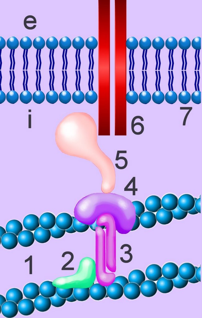
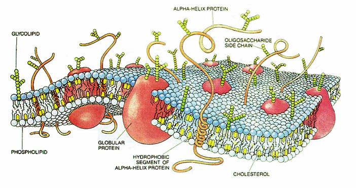
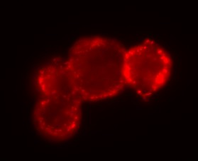
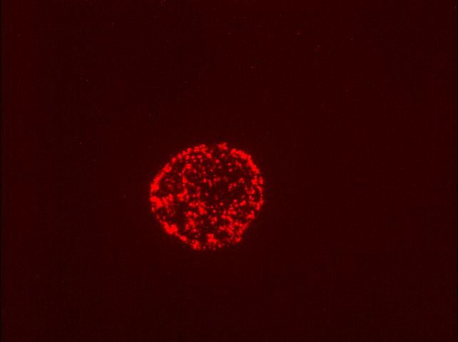
Cell membranes comprise phospholipids, sugars, and specialized proteins and perform a variety of functions such as adhesion, cellular communication, and maintenance of concentration gradients by active transport. These activities procede by means of specialized adhesion molecules, signaling molecules, ion channels and pumps, and receptor proteins.

The cell's cytoskeleton participates in chemotaxis, intracellular transport, and cell-to-cell adhesion. The cytoskeleton interacts with the extracellular environnment by way of transmembrane adhesion molecules (CAMs) and through surface protuberances such as cilia and flagellae (with various functions as mechanoreceptors, chemoreceptors, and the outer segment of the rods in the vertebrate retina). The centriolar components of the cytoskeleton organize the spindle apparatus on which the nuclear chromosomes translocate during mitosis and meiosis, phases of the reproductive cell cycle.

In multicellular organism, proliferation of cells must be balanced by cell death, usually through programmed, non-inflammatory apoptosis.

 Cell Adhesion Molecules  Immune Cytokines  Second Messengers  Cell signaling
 Apoptosis vs Necrosis  Apoptosis 

This site deals with Cell Biology, Cell Signaling, and the Molecular Biology of cells, particularly in relation to genetic mechanisms of biological evolution. Blue terms hyperlink to explanatory items so that you may navigate through items providing more detail. The site is searchable using the "Search this blog" box at top left. Use the "back" function to return to each departure item.

Alphabetic listing of Items - main items are bold
CELL BIOLOGY : A : active transport : adhesion : apoptosis : C : cell cycle : cell membranes : centrioles : (chemical gradients) : cilia and flagella : communication : concentration gradients : cytoplasm : cytoskeleton : D : death of cells : E : endocytosis : endoplasmic reticulum : endosomes : energy transducers : eukaryotic : exosome : F : flagella : G : Golgi apparatus : I : ion channels : L : lysosome : M : meiosis : microtubules : mitochondrion : mitosis : mitotic spindle : N : nuclear membrane : nuclear pore : nucleolus : nucleus : P : peroxisome : transport : phagocytosis : photosynthesis : physiological function : pinocytosis : plant cell : plasma membrane : prokaryotic : proteasome : protein degradation : protein pumps pumps : R : receptor proteins : receptor-mediated endocytosis : reproduction : ribosomes : RTKs : S : spindle : structure : V : vacuole : vesicle :

CELL SIGNALING : AKAPs : cellular signal transduction : concentration gradients : chemical gradients & communication : chemotaxis : cytokines : cytokine receptors : DAG : DAGKs : diacylglycerol : diacyl glycerol kinase : ERKs : GPCRs : GPCR families : hormones : neural action neural activity neuronal activity neuronal interconnections : neurotransmission : neurotransmitters : Nitric Oxide : PDZ domain : phosphorylation : phosphotransfer-mediated signaling pathways : PKA, protein kinase A : phospholipase C-gamma : PLC-G : PKC : protein kinase A : protein kinase C : protein tyrosine kinases protein kinases : Protein Kinase Signaling Networks : Ras : second messengers : signaling gradients (concentration gradients : chemical gradients) : signal transduction : two-component systems :

MOLECULAR BIOLOGY : amino acids : caspases : (concentration gradients) : (hormones) : proteins : Chemistry of Life site Biochemistry & Molecular Genetics : Molecular Genetics site

Items occur within Sections (listed in the sidebar). Items are listed in groups of 10 – to see more items, click on the lowest item in the sidebar. When visiting an item, the site title changes to purple – click on the title or “Home” to return to the main page.

The “Guide-Glossary” link below each item provides a glossary of terms ( # > 0 ), as a pop-up when reading within a Section, or as sub-script when visiting an Item.

| 0 Guide-Glossary links to this post

Inner Life of the Cell

For any who have not yet realized that the biology of cells can be fascinating – and aesthetic – Harvard University has sponsored an animation of the "Inner Life of the Cell" (HiRes, LoRes).


Unfortunately, the Internet-released version lacks a commentary soundtrack, settling instead for a music background that restricts the experience to pure aesthetics in the absence of explanation. My attempt at interpretation follows:

The animation opens with the rolling adhesion of a leukocyte within a blood vessel. The surfaces of two cells are shown adhering at contact points between adhesion molecules (selectin-saccharide).

We enter the cell and see a lipid raft, with its embedded sphingolipids and phosphatidyl choline(cholesterol+proteins), floating within the plasma membrane. Next we see a multi-protein focal contact, and then the cytoskeleton. After glancing back at the sub-plasma membrane 'geodesic' microfilaments, we pass down throught the cytoskeletal lattice and see actin microfilaments assembling, then depolymerizing after attachment of a protein (gelsolin?).

Next we see assembly and disassembly of tubulin. This interesting sequence is followed by my favorite segment, which is kinesin dragging an endosomal vesicle along a microtubule as kinesin-bigfoot 'walks' along the tubulin that is radiating from a centriole pair. A fellow kinesin, in the background, actively transports an endosome in the opposite direction.

Next, we approach the nuclear envelope with its embedded nuclear pore complexes. Several mRNA molecules with attached proteins exit the nucleus through the pores and assemble into loops within the cytoplasm, where the mRNA is scanned for a start codon, and is then translated into new polypeptide/protein chains by a ribosome. Globular proteins dimerize and tumble toward a mitochondrion.

Further translation injects a nascent protein chain through a pore into the endoplasmic reticulum as 'bigfoot' continues to clomp along tubulin, dragging a vesicle behind. Next, we see the Golgi apparatus budding vesicles before our plodding kinesin reaches the plasma membrane and proteins are released into the ECF by exocytosis. The newly synthesized integrins float on a lipid raft before unfolding into their active conformation, whereupon they snare a passing leukocyte and induce extravasation.

Happily, an extended version of the animation, complete with commentary, is now available on YouTube.


See a video of medical animator David Bolinsky describing the making of Inner Life of the Cell. More Cell Biology Videos.

The creationist mucusballs at the Dissembly Institute have plagiarized the footage, renamed it The Cell as an Automated City (presumably because that analogy is within the conceptual grasp of creationists), removed its credits, and are passing it off (implied) as Behe's research into the complexity 'created' by the IDer (aka God). The IDiots collect donations from credulous creationists as the DI Fellows pretend to conduct scientific investigation of IDiocy. I sincerely hope that Harvard University and XVIVO sue the pants of the IDiots, just as I'd like to see them tried and convicted of their plagiarism at the Kitzmiller vs Dover School Board trial.
See Creationist crooks pilfer Harvard's work . DI Fellows-- EXPELLED for plagiarism . Plagiarism and Intelligent Design .

Labels: , , , , , , , , , , , , , ,

| 0 Guide-Glossary links to this post

Fantastic Voyage

The following talk Fantastic voyage inside a cell was presented by medical animator Ted Bolinsky at TED, who describes the creation of Inner Life of the Cell. 9 min 57 secs.


Labels: , , , , ,

| 0 Guide-Glossary links to this post

Cell Biology Videos

On Cell Biology:
Inner Life of the Cell : Fantastic Voyage :

On Chemistry of Life:
transcription :

On YouTube:
The Inner Life of the Cell : Inner Life of a Cell: Leukocyte : Harvard Biovisions - The Inner Life of a Cell : Nano Visualization / Micro reality - Life Inside a Cell : Beginning of Life : CELL wrapping & DNA replication : CELL wrapping & DNA replication : THE MIRACLE OF MAN'S CREATION : Cell Signals (Part 2 of 2) : Sample clip from "Voyage Inside the Cell" V035 : The Life...of a cell. :

Labels: , , , , ,

| 0 Guide-Glossary links to this post

active transport

Active transport pumps require expenditure of energy, most often in the form of ATP, to transport hydrophilic macromolecules and ions across membranes against chemical or concentration gradients. The number of integral protein transporters in the membrane limits active transport.

Primary, or direct transport involves an energy expending conformational change in the membrane protein to transport a specific molecule or ion across the membrane. Secondary, or indirect transport utilizes energy directly to generate a transmembrane gradient down which ions move and then up or down which the coupled molecule or ion of interest is transported indirectly.

ATPases couple the hydrolysis of ATP (to ADP and Pi) with the transmembrane transport of ions against a concentration gradient. An example is Na+K+ ATPase, which pumps 3 Na+ ions out of the cell for 2 K+ ions it pumps into the cell. Because the pump moves ejects three Na+ for every two K+ moved inward, it generates a net electrical differential necessary for polarization. This electrical potential energy is essential for neuronal activity, and it supplies the energy needed for other types of transport such as symport and antiport. animation - Na K ATPase

Active transport of some substances against concentration gradients employs the ATPase-derived energy stored in ion gradients, such as proton (H+) or sodium (Na+) gradients, to drive transporter membrane proteins. In a symport, the ATPase transported molecule and the coupled, co-transported ion move in the same direction. Conversely, the ATPase transported molecule and the coupled, co-transported ion move in the opposite direction in an antiport.

Translocases utilize energy to move large molecules across the outer (Tom complex) and inner (Tim complex) mitochondrial membranes [image, more detail].

Џ beautiful Flash 8 animation - Inner Life of the Cell, which shows active transport of endosomes, and Interpretation: Inner Life of the Cell

Harvard University provides a wonderful video explaining operation of the F1-F0 ATPase. Џ

• A • adhesion • C • cell membranescellular signal transductioncentrioleschemotaxischloroplastciliacommunicationconcentration gradientscytokine receptorscytoplasmcytoskeleton • E • energy transducersendoplasmic reticulumendosomesexosome • G • Golgi apparatusGPCRs • H • hormones • I • ion channels • L • lysosome • M • meiosismicrotubulesmitosismitochondrion • N • Nitric Oxideneurotransmissionneuronal interconnectionsnuclear membranenuclear pore • P • pinocytosisproteasomepumps • R •receptor proteinsreceptor-mediated endocytosis • S • second messengerssignaling gradientssignal transductionspindlestructure • T • transporttwo-component systems • V • vacuolevesicle

animation - Na glucose symport : Carriers, Pumps, & Channels : The Virtual Cell Textbook - Cell Biology

Labels: , , , , , , , , ,

| 0 Guide-Glossary links to this post

adhesion

The cells of most eukaryotic species adhere to one another in co-operative multicellular organisms, whereas prokaryotes are unicellular organisms even though some species form colonies. Cell adhesion relies upon specialized transmembrane cellular adhesion molecules (CAMs) that usually extend from the intracellular space to the extracellular space where they may bind to other cell membranes or to the extracellular matrix. Within the intracellular domain, adhesion proteins adhere directly to, or are coupled to, the cell's cytoskeleton. Cadherin (calcium dependent adhesion) molecules are normally coupled by special linking proteins – catenins – to the cytoskeleton.

Intermediate filaments are about 10 nm diameter and provide tensile strength for the cell and connect adjacent cells through desmosomes (macula adherens).
cytoskeleton : diagram . desmosome : tem_desmosome : diagram . tight junction : diagram . gap junction : image - cytoskeleton : image_cytoskeleton : diagram - mechanism of ciliary motility Џ beautiful Flash 8 animation - Inner Life of the Cell, which shows the sequelae of adhesion-signaling, and Interpretation: Inner Life of the Cell Џ

 Cell Adhesion Molecules  Second Messengers  Cell signaling

There are several families of adhesion proteins, each with specific ligands of the same type (homophilic) or a different type (heterophilic).
1. integrins with heterophilic attachments to different (hetero) ligands in the extracellular matrix
2. selectins with heterophilic attachments to carbohydrate ligands
3. Ig superfamily proteins with selectins with heterophilic attachments to: a) integrin ligands, and b) Ig superfamily proteins of a different (hetero) type, and
homophilic attachments to the Ig superfamily proteins of the same (homo) type
4. cadherins with homophilic attachments to cadherins of the same type, or by way of catenins (right - click to enlarge, description), to the cytoskeleton

Cell adhesion is important in:
1. maintaining contact within solid tissue
2. embryogenesis (morphogenesis)
3. migration of single cells such as leukocytes within multicellular organisms
4. maintaining contact between neuronal elements
5. virulence of virions and bacteria

Some signaling molecules act as adhesion receptors, and cluster in focal adhesions upon ligand binding. (Rho protein). A variety of integrins, which are transmembrane heterodimeric adhesion receptors are known to support adhesion-dependent growth factor-activation of MAP kinase. Focal adhesions are rich in tyrosine phosphorylated proteins, coupling cell adhesion to signal transduction pathways in the cell. Various adhesion receptors, such as integrin, are closely linked to protein kinases and phosphatases. Grb2 links focal adhesion kinase (FAK) to the Ras pathway when Grb2 is phosphorylated after binding to FAK. The 85 kDa subunit of the PI 3-kinase is also phosphorylated after binding to FAK. Thus, FAK is a key component in the assembly of focal contact structures that influence cytoskeletal organization and signal transduction.

Engagement of ICAM-1, a member of the immunoglobulin supergene family (Ig), has been documented to activate specific kinases through phosphorylation, resulting in activation of transcription factors, increased cytokine production, increased cell membrane protein expression, production of reactive oxygen species, and cell proliferation.

cadherins : Ig superfamily proteins : integrins : selectins : ICAMs :  Cell Adhesion Molecules
 Second Messengers  Cell signaling .

Cells form a variety of intermembranous junctions: adherens junctions, desmosomes (plasmodesmata), focal contacts, gap junctions, hemidesmosomes, tight junctions.

cytoskeleton : diagram . desmosome : tem_desmosome : diagram . tight junction : diagram . gap junction : image - cytoskeleton : image_cytoskeleton : diagram - mechanism of ciliary motility :

• A • adhesion • C • cell membranescellular adhesion moleculescellular signal transductioncentrioleschemotaxischloroplastcilia & flagellacommunicationconcentration gradientscytokine receptorscytoplasmcytoskeleton • E • energy transducersendoplasmic reticulumendosomesexosome • F • flagella & cilia • G • Golgi apparatusGPCRs • H • hormones • I • ion channels • L • lysosome • M • meiosismicrotubulesmitosismitochondrion • N • Nitric Oxideneurotransmissionneuronal interconnectionsnuclear membranenuclear pore • P • pinocytosisproteasomepumps • R • receptor proteinsreceptor-mediated endocytosis • S • second messengerssignaling gradientssignal transductionspindlestructure • T • transporttwo-component systems • V • vacuolevesicle

| 0 Guide-Glossary links to this post

apoptosis

Cellular death-by-suicide is part of normal development, and is termed apoptosis or programmed cell death (PCD). Cysteine Aspartate Specific ProteASEs – caspases – are active in apoptosis, as are p53, a tumor suppressor gene, and FAS gene, which is member 6 of the tumor necrosis factor receptor superfamily (TNF). In contrast to apoptosis, necrosis is cell death that results from cytotoxic, injurious stresses that are too severe for correction by the cellular stress response.

▼: AIF : ANT : apoptotic appearance : apoptosomes : apoptotic bodies : Bcl-2 : Bcl-2 family : caspase cascadecaspases : cytochrome C : death domain : death receptor pathway : DISC : Endonuclease G : FADD : FAS gene : homeostasis and apoptosis : MBR : mitochondrial pathway : necrosis : PBR : porin : proteolytic cascade : PT pore : TNF-R : tumor suppressors : Smac/DIABLO : VDAC : ▼

Tumor suppressor proteins such as p53 act as cell-cycle repressors and/or promoters of apoptosis through:
1. interruption of cell cycle, preventing cell division,
2. halting the cell cycle if DNA damage is not yet repaired,
3. inducing apoptosis if DNA damage cannot be repaired,
4. promoting cell adhesion and contact inhibition, which prevent invasion and metastasis.

Both the death receptor pathway and the mitochondrial pathway lead to the activation of an initiator caspase, which initiates a proteolytic cascade, ultimately leading to apoptotic cell death.

Necrosis results from impairment of the cell’s ability to maintain homeostasis due to breakdown of the plasma membrane. This leads to influx of water as pumps fail and osmotic gradients reverse. Ultimately organelles (especially mitochondria) swell and hydrolytic, lysosomal enzymes are released into the cytosol, leading to cellular swelling and rupture (lysis). So, in vivo, necrotic cell death is often associated with extensive tissue damage resulting in an intense inflammatory response.

Apoptosis, by contrast, occurs under normal physiological conditions when the cells respond to signals as active participant in their own deaths. Apoptosis is a part of normal cell turnover and tissue homeostasis. It plays a significant role in embryogenesis, induction and maintenance of immune tolerance, development of the nervous system, and atrophy of endocrine-dependent tissue.

[] Artist's impression apoptosis []

Cells undergoing apoptosis show characteristic morphological and biochemical features, which include chromosomal changes: chromatin aggregation, nuclear and cytoplasmic condensation (2), and partition of cytoplasm and nucleus into apoptotic bodies (apoptosomes). These are membrane bound-vesicles that contain ribosomes and intact mitochondria and nuclear material (3). Apoptotic bodies are rapidly recognized in vivo and and are phagocytozed by either macrophages or adjacent epithelial cells (4), without inflammatory response. In vitro, apoptotic bodies and remaining cell fragments ultimately swell and lyse (“secondary necrosis"). [more] Џ apoptosis animations Џ

Tables  Apoptosis vs Necrosis  Apoptosis 

Death receptor pathway:
Death receptors include the TNF-R (tumour necrosis factor receptors) and CD95 (Apo-1 or Fas) receptor families. Ligands for these death receptors are termed TNF-α and CD-95L (FasL), respectively. TNF-α is a highly cytotoxic molecule and the TNF-R1 receptor is widespread, with the result that TNFα has a low tolerated dose. Several members of the TNF-R1 family share a homologous region known as the death domain (DD), which is a protein-protein interaction domain that binds to adaptor proteins such as the adaptor protein FADD (Fas-Associated Death Domain). The Death-Inducing Signalling Complex (DISC) comprises death receptor ligands, death receptors and adaptor proteins such as FADD. Activation of death receptors by binding of ligands such as CD-95L (FasL) and TNF-α leads to caspase-8 activity.

Mitochondrial pathway:
Mitochondria play a central role in apoptosis and display an increase in mitochondrial membrane permeability during apoptosis. Mitochondria participate in apoptosis, and may be necessary for induction of apoptosis by apoptotic stimuli such as DNA damage. Pro-apoptotic and anti-apoptotic members of the Bcl-2 family are believed to regulate the release, through the mitochondrial PT pore, of pro-apoptotic substances such as AIF, Endonuclease G, Smac/DIABLO and cytochrome C. Mitochondrial efflux of cytochrome-c drives generation of the apoptosome (apoptotic body) in the cytoplasm. This in turn leads to caspase-9 activity.

Bcl-2 family: Members of the Bcl-2 subfamily (Bcl-2, Bcl-xL, Bcl-w) are localized on the outer mitochondrial membrane and show anti-apoptotic activity. They possess all four BH (Bcl-2 Homology) domains (BH1 to BH4). (Anti-apoptotic members of the Bcl-2 family have all four BH domains, while pro-apoptotic members have less BH domains.) Bax subfamily (Bax, Bak, BAD) : Bax is Bcl-2 Associated X protein, a pro-apoptotic member of the Bcl-2 family, which contains BH1 to BH3. BH3 only subfamily: Bid or tBid is (truncated) BH3-Interacting Domain death agonist, is a soluble, pro-apoptotic member of the Bcl-2 family, which contains only the BH3 domain.

Pro-apoptotic proteins : Bad, Bax, Bid, Bak, Bik, Bim, Bmf, Bok, Puma.
Anti-apoptotic proteins : Bcl-2, Bcl-XL, Mcl-1, Bcl-w

Released through the Mitochondrial Permeability Transition Pore (PT pore) are:
1. Apoptosis Inducing Factor (AIF) is a flavoprotein that is translocated to the nucleus where it causes DNA fragmentation into fragments of about 50kb.
2. Endonuclease G (Endo-G) is translocated to the nucleus, where it degrades single stranded DNA.
3. Smac/DIABLO is "Second Mitochondrial Activator of Caspases/Direct IAP Binding protein with Low pI", which inhibits IAP activity, and is therefore pro-apoptotic.
4. Cytochrome c is an essential component for the transportation of electrons during mitochondrial oxidative phosporylation. When released from the mitochondria, cytochrome c drives the formation of the apoptotic body (apoptosome).[s, Refs]

The PT pore is constituted of ANT, PBR (MBR), and VDAC (porin), and the mitochondrail membrane porosity is modified by Bcl-2 regulators.

Tables  Apoptosis vs Necrosis  Apoptosis  Џ beautiful Flash 8 animation - Inner Life of the Cell : and Interpretation: Inner Life of the Cell Џ

• A • adhesion • C • cell membranescellular adhesion moleculescellular signal transductioncentrioleschemotaxischloroplastcilia & flagellacommunicationconcentration gradientscytokine receptorscytoplasmcytoskeleton • E • energy transducersendoplasmic reticulumendosomesexosome • F • flagella & cilia • G • Golgi apparatusGPCRs • H • hormones • I • ion channels • L • lysosome • M • meiosismicrotubulesmitosismitochondrion • N • Nitric Oxideneurotransmissionneuronal interconnectionsnuclear membranenuclear pore • P • pinocytosisproteasomeprotein degradationpumps • R • receptor proteinsreceptor-mediated endocytosis • S • second messengerssignaling gradientssignal transductionspindlestructure • T • transporttwo-component systems • U • ubiquitin • V • vacuolevesicle

Џ Quicktime video apoptosis : Apoptosis and Caspase :

Genome Biology Full text DNA-damage signaling and apoptosis: "Cytochrome c binds the apoptosis-activating factor 1 (Apaf1) protein, leading to oligomerization of Apaf1 and caspase 9 into a large 'apoptasome', which then initiates a cascade of caspase activation. Although some non-caspase targets of caspase activation are known, the consequences of proteolysis of these targets are not well understood. Similarly, the events upstream of activation of the caspase cascade in response to DNA damage are not well known; in particular, it is not clear what regulates the decision to undergo apoptosis or to arrest cell proliferation and repair the damage."

apoptosis: Bcl-2 proteins
Apoptosis - Bcl-2 proteins: "The bcl-2 proteins are a family of proteins involved in the response to apoptosis. Some of these proteins (such as bcl-2 and bcl-XL) are anti-apoptotic, while others (such as Bad or Bax) are pro-apoptotic . The sensitivity of cells to apoptotic stimuli can depend on the balance of pro- and anti-apoptotic bcl-2 proteins. When there is an excess of pro-apoptotic proteins the cells are more sensitive to apoptosis, when there is an excess of anti-apoptotic proteins the cells will tend to be less sensitive."

Џ apoptosis animations : Kimball's Apoptosis Page : Apoptosis - Website : Apoptosis Website : Wikipedia on apoptosis : pathology : Cell Suicide in Health and Disease : Google apoptosis Џ animation - zeiosis Џ Apoptosis, Bcl2, Mitochondria~click on Q for animation Џ Apoptosis~time-lapse movie : art~apoptosome formation : art~inactivation of DNA repair enzymes : Cell Death Pathways - diagram

▲: AIF : ANT : apoptotic appearance : apoptosomes : apoptotic bodies : Bax : Bcl-2 : Bcl-2 family : caspase cascadecaspasescellular stress response : cytochrome Cdeath of cells : death domain : death receptor pathway : DISC : Endonuclease G : FADD : Fas geneFAS gene : homeostasis and apoptosis : MBR : mitochondrial pathwaymitochondria : necrosis : PBR : porin : proteolytic cascade : PT pore : TNF-RTNF-R : tumor suppressors ¤ tumor suppressors : Smac/DIABLO : VDAC : ▲

Top

Labels: , , , , , , , , , , , , , , ,

| 0 Guide-Glossary links to this post

cell growth

The term cell growth can refer to an increase in cellular size or to cellular doubling by reproduction (proliferation).

Cell growth is normally stimulated by growth factors and mitogens, regulated by signaling pathways, and balanced by apoptosis.

Џ beautiful Flash 8 animation - Inner Life of the Cell : and Interpretation: Inner Life of the Cell Џ

| 0 Guide-Glossary links to this post

cell membranes

Cell membranes provide adjustable barriers between the cell and the extracellular environment (ESF) or adjacent cells in eukaryotes. The membranes of cellular organelles provide functional compartmentalization from the cytosol.

The cell itself is surrounded by the plasma membrane, and specific functional membranes form intracellular organelles (endoplasmic reticulum, Golgi apparatus) or isolate the contents of cellular organelles (chloroplasts, endosomes, exosomes, lysosomes, mitochondria, peroxisomes) from the cytoplasm. art - cell membrane translucent : art - cell membrane opaque :

Left -click to enlarge image: Schematic three dimensional cross section of a cell membrane. There are two major components of this dynamic, fluid, structure: lipids and proteins. The lipid bilayer provides the basic structure within which proteins are free to diffuse. Sugar moieties can be present as part of either proteins (glycoproteins) or of lipids (glycolipids). Cholesterol intercalates between lipid molecules and affects membrane fluidity/stability.

Cell membranes are variably constituted of carbohydrates (adhesion and address loci), phospolipid bilayers (hydrophobic barriers), and proteins, which control permeability and cellular signalling. Peripheral membrane proteins are confined to the surfaces of membranes while integral membrane proteins are embedded in the membrane and may pass through the lipid bilayer one or more times.

Specialized membrane proteins function in cell adhesion (junctions) and as energy transducers, enzymes, ion channels, pumps, and receptors for neurotransmitters and hormones. Cell junctions utilize proteins that anchor cells together (desmosomes), that occlude passage of water between cells (tight junctions), and that permit direct communication between cells (gap junctions).

Lipid rafts are mobile areas within cell membranes that are more rigid than the rest of the bilayer by virtue of their enrichment in different lipids, cholesterol, and proteins. []fm, fm2[]Several types of lipid raft have been postulated: inside rafts (PIP2 rich and caveolae) and outside rafts (GEM) constituted of the three postulated compositions: caveolae (with caveolin-1), glycosphingolipid enriched membranes (GEM), and polyphosphoinositol rich rafts. Caveolin-1 is a 21kDa cholesterol binding, integral membrane protein. Muscle-expressed caveolin-3, which is involved in some types of muscular dystrophy, forms muscle-type caveolae.

Glycosphingolipids, and other lipids with long, straight acyl chains are preferentially incorporated into the rafts such that fatty-acid chains tend to be extended and thus more tightly packed, creating higher order domains. It is believed that rafts exist in a separate ordered phase that floats within the regular sea of poorly ordered lipids.

It is most likely that lipid rafts are involved in cellular signaling. Many actin binding proteins (ABP) bind to, and are regulated by polyphosphoinositides. These ABPs include gelsolin, which is a Ca2+, pH and polyphosphoinositide-regulated actin capping/severing protein that partitions neural membranes into biochemically isolated rafts. []fluorescence micrograph gelsolin[] GEMs also appear to link to the actin cytoskeleton through ABPs, in particular ERM proteins through EBP50, which binds members of the ERM proteins through the ERM C-terminus.

Џ beautiful Flash 8 animation - Inner Life of the Cell, which shows plasma and organellar membranes, a lipid raft, and exocytosis; and, Interpretation: Inner Life of the Cell Џ

• A • adhesion • C • cell membranescellular signal transductioncentrioleschemotaxischloroplastciliacommunicationconcentration gradientscytokine receptorscytoplasmcytoskeleton • E • energy transducersendoplasmic reticulumendosomesexosome • G • Golgi apparatusGPCRs • H • hormones • I • ion channels • L • lysosome • M • meiosismicrotubulesmitosismitochondrion • N • Nitric Oxideneurotransmissionneuronal interconnectionsnuclear membranenuclear pore • P • pinocytosisproteasomepumps • R •receptor proteinsreceptor-mediated endocytosis • S • second messengerssignaling gradientssignal transductionspindlestructure • T • transporttwo-component systems • V • vacuolevesicle

diagram . desmosome : tem_desmosome : art - tight junction zonula adherens desmosome gap : diagram . tight junction : diagram . gap junctionball-stick - globular proteins in phospholipid bilayer : ball-stick - carrier proteins : ball-stick - marker protein : ball-stick - marker proteins : ball-stick - receptor proteins : ball-stick - ion channel proteins: animation - carrier proteins : animation - receptor protein : animation - cholesterol

| 0 Guide-Glossary links to this post

centrioles

Centrioles organize the spindle apparatus on which the chromosomes move during mitosis. Cilia and flagella are organized from peripheral centrioles (basal bodies).

Centrioles consist of 9 sets of triplet microtubules, and centrioles are arranged in pairs perpendicular to each other (tem - 9 triplet pair). animation - spinning centriole pair : tour centriole : zoom in on centriole. Џ beautiful Flash 8 animation - Inner Life of the Cell, which shows a centriole pair with radiating microtubules, and Interpretation: Inner Life of the Cell Џ

Unlike cilia and flagella, which are organized from microtubule organizing centers (basal bodies) at the cell periphery, centrioles have no central doublet of microtubules. Centrioles replicate autonomously, beginning from centers that contain proteins needed for their formation (tubulin, etc.). Procentrioles form first, each erecting a single microtubule from which the triplet can form (diagram - centriole formation). After a single centriole is constructed, daughter centrioles grow out from the tubules at right angles. In a non-dividing cell, they move to the periphery to form the basal body for the cilium (tem - basal bodies). In a dividing cell, the second centriole moves to the daughter cell (in a dividing cell). Where spindles are essential for chromosomal separation during reproduction, cilia are essential for cellular differentiation during embryologic development.

The microtubule organizing center, also called a basal body, lies at the base of the cilium. tem - basal bodies. The basal body is created as the centriole (functioning as a microtubular structure essential to cell division) migrates to the surface. The transition zone between axoneme and basal body serves as a docking station for intraflagellar transport and motor proteins. During intraflagellar transport (IFT) materials needed to build the cilia are carried to the ciliary tip and spent materials are carried down to the ciliary body.

More: Cilia, Flagella, and Centrioles : Cilia and flagella : HHMI Bulletin September 2005: The Importance of Being Cilia : Google cilia : Virtual Cell Textbook - Cell Biology :

| 0 Guide-Glossary links to this post

chemotaxis

Chemotaxis is a signal transduction system that confers to the ability of cells to respond to spatial and temporal chemical gradients. In prokaryotes and unicellular eukaryotes, chemotaxis controls motile behavior. In multicellular eukaryotes, chemotaxis plays an important role in determining the migration of cells in many biological processes including immune response, development, embryogenesis, wound healing, and angiogenesis (1, 2, 3, 4). During embryogenesis, cells respond to chemotactic stimuli, enabling the organization of tissues and organs, and neuronal migration (5, 6, 7). An elaborate network of chemoattractants directs leukocytes to their correct locations and facilitates cell-cell interactions in the immune system. Chemotaxis is central to wound healing, and operates in disease states such as metastasis and atherosclerosis (8, 9, 10, 11, 12).

Even though the motility apparatus differs among organisms, the general mechanism of control of chemotaxis is conserved throughout all prokaryotic bacteria and archaea. Central to chemotactic control is the two-component system in which phosphorylation of a response regulator reflects phosphorylation of a histidine autokinase that senses environmental parameters (117). This is the commonest mode of signal transduction system in bacteria, and the two component system controls diverse processes such as gene expression, sporulation, and chemotaxis.

Prokaryotic chemotaxis proteins comprise four groups—a signal recognition and transduction group, an excitation group, an adaptation group, and a signal removal group (to dephosphorylate CheY-P). The signal recognition and transduction group includes the receptors and ligand binding proteins, which are capable of binding effectors outside the cell; a few receptors, however, are cytoplasmic: (image general chemotaxis model : Table proteins in chemotaxis ). Prokaryotes react as point sensors, determining static gradients by temporal sampling.

The mechanism of chemotaxis in eukaryotic cells is different than that in prokaryotes, though eukaryotic cells also respond to chemical, signaling gradients. By virtue of their greater size, eukaryotes are able to detect concentration gradients along their plasma membranes where receptor occupancy differences provide input for discrepancy sensing of gradients. In response to chemoattractant gradients, the imbalance between activator action and inactivator action results in a spatially oriented persistent signaling. Most eukaryotic cells employ GPCRs to detect these chemical signals, so signaling is amplified by a substrate supply-based positive feedback that acts through small G-proteins. This amplification is activated only in the continuous presence of the external signal gradient, thus providing the mechanism for sensitivity to gradient alterations. Other membrane receptor include amino acids, insulin, and vasoactive peptides. Chemotactic receptors are triggered by formyl peptides, chemokines (α, CXC; β, CC; δ, CX3C; γ, C), and leukotrienes (autocrine and paracrine eicosanoid lipid mediators derived from arachidonic acid by 5-lipoxygenase).

Following signal detection by a GPCR, a locally acting activator (PI3-kinase) and a globally acting inactivator (PTEN or a similar phosphatase) are coordinately controlled by the G-protein activation [1]. The signaling system adapts to spatially homogeneous changes in the chemoattractant. In chemoattractant gradients, an imbalance between the action of the activator and the inactivator results in a spatially oriented persistent signaling, amplified by a substrate supply-based positive feedback acting through small G-proteins. The amplification is activated only in a continuous presence of the external signal gradient, thus providing the mechanism for sensitivity to gradient alterations.

During the course of evolution, eukaryotic cilia have been adapted to function as mechanoreceptors and chemoreceptors. Eukaryotic cilia and flagella differ from the flagella of eubacteria, which differ in turn from the flagella of archaebacteria. Organisms such as Amoebae and Tetrahymena are able to creep or swim, utilizing the cytoskeleton/cilia/flagella. The slime mold, Dictyostelium discoideum commences gradient-sensing with modification of the phospholipid membrane at the cell's leading edge. The modification includes the recruitment of various proteins to the membrane, initiating a process that culminates in pseudopod extension in the direction of the chemoattractant. Cells exposed to shallow chemoattractant gradients respond with strong accumulation of the enzyme phosphatidylinositol 3-kinase (PI3-K, PI3K) and its D3-phosphoinositide product (PIP3) at the plasma membrane exposed to the highest chemoattractant concentration, whereas PIP3-degrading enzyme PTEN and its product phosphatidylinositol bisphosphate (PIP2) localize in a complementary pattern [Gamba]. This early symmetry-breaking event is a mandatory step for directed cell movement elicited by chemoattractants. It has been suggested that 'directional sensing is the consequence of a phase-ordering process mediated by phosphoinositide diffusion and driven by the distribution of chemotactic signal'.

When starved, D. discoidem cells differentiate, polarize, and migrate directionally toward secreted 3',5'-cyclic adenosine monophosphate (cAMP). The cAMP is detected by four GPCRs, designated cAR1–cAR4, which are coupled to a single heterotrimeric G-protein (19). Mammalian leukocytes couple 20 types of chemoattractant, chemokine receptors to the same G-protein, Gi (20, 21). Mammalian systems also employ chemoattractant-elicited transient increases in phosphoinositides (PIs),1 cAMP, cGMP, inositol trisphosphate (IP3), and Ca2+, and rearrangements in the cytoskeleton (16, 22). The PI3-kinase elevates local levels of PIP3, which determines the sites of new actin-filled projections. PIP3 is an important intermediate in chemotactic signaling in D. discoideum, amoebae, and mammalian leukocytes (23, 24, 25, 26, 27, 28, 29, 30, 31, 32, 33, 34). 'Robust control of the temporal and spatial levels of PIP3 is achieved by reciprocal regulation of PI3K and PTEN.'[2, fft]

Ras, PI3K, and TOR are known as key regulators of cellular growth, and play a critical role in regulating the actin cytoskeleton, cell polarity, and cellular movement, indicating that multiple steps in the signal transduction pathway coordinately regulate cell motility.

Chemotaxis is important early in embryologic development and in the immune system. Within multicellular organisms, specific types of cells such as those of the immune/hematologic system (granulocytes, monocytes, macrophages, lymphocytes) and specialized, independent cells (mast cells, endothelial cells, fibroblasts, and, unfortunately, tumor cells) are capable of motile responses to chemical signals. Motility and migration employ cellular adhesion molecules.

Џ beautiful Flash 8 animation - Inner Life of the Cell, which shows chemotaxis in action, and Interpretation of Inner Life of the Cell Џ


· adenylyl (adenylate) cyclase · calcium ions · cAMP-dependent protein kinase · CDKs · cyclin-dependent kinases · DAG · diacylglycerol · DNA ligases · ERKs · GPCRs · GPCR families · guanylate cyclases · guanyl cyclase · inositol triphosphate · IP3 · MAP kinases · mitogen activated protein kinases · phosphatases · phosphodiesterases · phospolipases · phosphorylation · PKA · PKC · phospholipase C-gamma · protein kinase A · protein kinase C · protein tyrosine kinases (PTKs) · receptor tyrosine kinases · second messengers · second messenger cAMP · second messenger cGMP · signal transduction · two-component systems ·

Tables  Cell signaling  Cell Adhesion Molecules  Immune Cytokines  Second Messengers Receptor Tyrosine Kinases(RTK)  Second Messengers  Phosphate-handling Enzymes 

: confocal image -GFB amoeba chemotaxis : : Links to videos of Dictyostelium: Cytokinesis : Cytokinesis Defects : Chemotaxis : Development : Morphogenesis : Motility : Phagocytosis : Videos Chemotaxis : Chemoattractant receptors in chemotaxis : G-protein b-subunits in chemotaxis : PH-domain translocation with uniform stimulus : PH-domain translocation during chemotaxis : PH-domain translocation in immobilized cells : PH-domain dynamics in immobilized cells : Digitized wild-type cell in chemotaxis : Digitized clathrin heavy chain null cell in chemotaxis :

In addition to chemotaxis, cells perform:
1) chemokinesis – scanning the environment,
2) haptotaxis – generation of a chemoattractant gradient in the ECM employed in transendothelial migration and angiogenesis, and
3) necrotaxis – negative or positive response to chemoattractants released from apoptotic or necrotic cells.

• A • adhesion • C • cell membranescellular adhesion moleculescellular signal transductioncentrioleschemotaxischloroplastcilia & flagellacommunicationconcentration gradientscytokine receptorscytoplasmcytoskeleton • E • energy transducersendoplasmic reticulumendosomesexosome • F • flagella & cilia • G • Golgi apparatusGPCRs • H • hormones • I • ion channels • L • lysosome • M • meiosismicrotubulesmitosismitochondrion • N • Nitric Oxideneurotransmissionneuronal interconnectionsnuclear membranenuclear pore • P • pinocytosisproteasomepumps • R • receptor proteinsreceptor-mediated endocytosis • S • second messengerssignaling gradientssignal transductionspindlestructure • T • transporttwo-component systems • V • vacuolevesicle

| 0 Guide-Glossary links to this post

chloroplast

The chloroplast is the site of photosynthesis in eukaryotic cells, and is the site of the Calvin cycle just as the mitochondrion is the site of oxidative phosphorylation.

The thylakoid membrane, with its embedded photosystems, is the structural unit of photosynthesis. Both photosynthetic prokaryotes and eukaryotes possess membranes with embedded photosynthetic pigments. Only eukaryotes, which have a nuclear membrane and membrane-enclosed organelles, have chloroplasts with an encapsulating membrane. The chloroplast has three compartments, while the mitochondrion has only two. Compartments within a chloroplast are the intermembranous space [3], the stroma [6], and the thylakoid lumen (8) within stromal and granal thylokoids [4,5].

1. outer membrane
2. inner membrane
3. intermembranous space
4. stromal thylakoid
5. granal thylakoid
6. stroma (cytosol)
7. granum (a stack of thylakoids)
8. internal lumen of granal and stromal thylakoids
(click to enlarge image)


The typical higher plant chloroplast is lenticular and approximately 5 microns at its largest dimension. Plant cells contain from 1 to 100 chloroplasts, depending on the type of cell. The mature chloroplast is typically bounded by inner and outer membranes that possess significantly different chemical constituents. (tem - chloroplast & microbodies, tem - chloroplast, micro - chloroplast) In addition to enzymes that function in photosynthesis, chloroplasts also contain a circular DNA molecule (cpDNA below) and the protein-synthetic machinery characteristic of prokaryotes.

Each chloroplast contains about 40 to 80 grana (7), and each grana comprises about 5 to 30 thylakoids. The thylakoids are membranous disks about .25 to .8 microns in diameter, which contain protein complexes, pigments, and other accessory components. The phospholipid bilayer of the thylakoid is folded repeatedly into stacks of grana. (details) These stacks are connect by channels to form a single functional compartment.

The smooth outer membrane (1) is freely permeable to molecules, and resembles the chemical constitution of the eukaryotic plasma membrane. The smooth inner membrane (2) contains many integral transporter proteins that regulate the passage of small molecules like sugars, and proteins (synthesized in the cytoplasm of the cell, but utilized within the chloroplast). The inner membrane chemically resembles prokaryotic cell membranes.

The thylakoid is the site of oxygenic photosynthesis in eukaryotic plants and algae, and in prokaryotic Cyanobacteria. Cyanobacteria possess thylakoid membranes, but as prokaryotes they do not contain chloroplasts. Chlorophyll, accessory pigments, and other integral membrane proteins transduce light energy to provide excited electrons (excitons) to electron transport chains, powering the formation of NADPH and ATP during photophosphorylation.

The folded thylakoid membranes perform the light reactions of photosynthesis utilizing Photosystems I and II, both of which include chlorophyll and carotenoid molecules (bsim - chlorophyll, spfim - chlorophyll, bsim - carotenoid). The reaction center chlorophyll molecule within the antenna of photosystem I responds most strongly to 700 nm light, and is therefore termed P700. The reaction center within the antenna of photosystem II responds most to 680 nm light, and is accordingly called P680.

Photosystem I evolved very early, and it is found in nonoxygenic phototrophs; photosystem II evolved later. Because the PSII photosystem is most sensitive to shorter wavelength 680 nm light, it absorbs slightly more energy than the P700-PSI system.

The electron transport system of each photosystem is embedded within the thylakoid membrane and functions in the production of ATP. The system comprises membrane-bound electron carriers that pass electrons from one molecule to the next. The purple bacteria utilize only one photosystem (PSI), while oxygenic phototrophs utilize two photosystems (PSI and PSII). Prokaryotes contain bacteriochlorophylls, which differ both chemically and in absortption spectra from those of Cyanobacteria and chloroplasts. Chemical differences involve the phytol side chain, groups attached to the porhyrin ring, and the saturation of one pyrrole subunit of the porphyrin ring. Green bacteria possess highly efficient membrane-bound chlorosomes.The photosynthetic machinery of nonoxygenic photosynthetic purple bacteria is often located in intracytoplasmic membranes. It is not yet known whether or not these membranes are similar to the thylakoid membrane of oxygenic phototrophs, or whether these intracytoplasmic membranes of nonoxygenic phototrophs are merely an extension of the plasma membrane.

As intracellular plant organelles, chloroplasts are classified as plastids. Chloroplasts originate within the eukaryotic photosynthetic cell either by division of pre-existing plastids or from protoplastids (proplastid). These proplastids are organelles with little internal structure, enclosed within two dissimilar membranes. It is assumed that thylakoid membranes formed during the chloroplast maturation process and are derived from the inner membrane of the proplastid and chloroplast. diag - chloroplast development

The current consensus is that chloroplasts originated from Cyanobacteria that have become endosymbionts. This is an origin analogous to the endosymbiotic origin of mitochondria, which are believed derived from "purple bacteria", alpha-proteobacteria most closely related to Rickettsiales.

The cpDNA genes encode some of the molecules needed for chloroplast function. Hundreds of others are transcribed from genes in cellular nucleus, translated into proteins in the cytoplasm, and transported into the chloroplast. Thus, the majority of the proteins expressed in the plastid are encoded in the nuclear genome of the host cell. This genetic dependency on the cellular genome distinguishes organelles from obligate endosymbionts. Gene loss, substitution of nuclear genes, and gene transfer cause reduction in the size of the plastid genome (see Endosymbiotic Gene Transfer).

Chloroplast RNA-binding and pentatricopeptide repeat proteins:
Chloroplast gene expression is mainly regulated at the post-transcriptional level by numerous nuclear-encoded RNA-binding protein factors. In the present study, we focus on two RNA-binding proteins: cpRNP (chloroplast ribonucleoprotein) and PPR (pentatricopeptide repeat) protein. These are suggested to be major contributors to chloroplast RNA metabolism. Tobacco cpRNPs are composed of five different proteins containing two RNA-recognition motifs and an acidic N-terminal domain. The cpRNPs are abundant proteins and form heterogeneous complexes with most ribosome-free mRNAs and the precursors of tRNAs in the stroma. The complexes could function as platforms for various RNA-processing events in chloroplasts. It has been demonstrated that cpRNPs contribute to RNA stabilization, 3´-end formation and editing. The PPR proteins occur as a superfamily only in the higher plant species. They are predicted to be involved in RNA/DNA metabolism in chloroplasts or mitochondria. Nuclear-encoded HCF152 is a chloroplast-localized protein that usually has 12 PPR motifs. The null mutant of Arabidopsis, hcf152, is impaired in the 5´-end processing and splicing of petB transcripts. HCF152 binds the petB exon–intron junctions with high affinity. The number of PPR motifs controls its affinity and specificity for RNA. It has been suggested that each of the highly variable PPR proteins is a gene-specific regulator of plant organellar RNA metabolism.
T. Nakamura, G. Schuster, M. Sugiura and M. Sugita Chloroplast RNA-binding and pentatricopeptide repeat proteins Biochem. Soc. Trans.. (2004) 32, (571–574)

External link Chloroplast : micro - chloroplast : tem - chloroplast & microbodies : tem - chloroplast : tem - mitochondrion : diag - chloroplast development : diag - plant cell constituents : photochemistry diag - Calvin Benson , image - light reactions,

| 0 Guide-Glossary links to this post

chromosomes

Genetic archival material is condensed into chromosomes. (Left - click to enlarge image)

1. one of two sister chromatids
2. centromere (kinetochore region)
3. short p arm
4. long q arm

A chromosome contains a long strand of deoxyribonucleic acid containing genes, regulatory sequences, and non-coding sequences of nucleotides, in association with proteins. The full chromosomal complement of a cell comprises the genome, which is the complete hereditary information of an organism contained within macromolecules of archival DNA.

The multiple nuclear chromosomes of eukaryotes exist as nucleosomes in which long helical strands of DNA are wrapped around structural proteins called histones – this composite material is termed chromatin (diagram). Each eukaryotic chromosome comprises one or two (sister) chromatids (1), each with a kinetochore (2) for attachment to a microtubule of the spindle apparatus during cell division. Chromatids have a long (q) and a short (p) arm attached to the centromere (2). Sister chromatids attach to each other, or to the spindle apparatus, by means of special proteins and DNA base sequences in the kinetochore region.

Chromosomes (Gk. 'colored bodies') are most visible during metaphase (tem), and least condensed (dispersed) when participating in expression (tem, tem2) such as occurs in cells with large undifferentiated nuclei (colored tem cancer cell, fluorescence microscopy of cancer, stem cells, immature cell with oncogene (black dots) ).

Prokaryotes mostly possess one, sometimes two* chromosomes termed nucleoids (tem). Prokaryotes lack a membrane enclosed nucleus and their DNA is usually contained in circular structures located within the cytosol, but may be organized as linear strands that are typically attached to the plasma membrane. Plasmids are small circular, extrachromosomal genetic elements that can be transmitted from one bacterium to another through pili during conjugation. (more)

Chromatin (DNA plus histone protein) exists in two basic forms (tem):
1. Euchromatin, from which DNA is being actively transcribed (expressed) into RNA for ultimate translation into polypeptide and protein molecules.
2. Heterochromatin, which consists of either:
a. Facultative heterochromatin, which is sometimes expressed.
b. Constitutive heterochromatin, which is located around the centromere and usually contains repetitive sequences, and which is never expressed.

Nucleosomes compact the DNA and render it inaccessible and hence inactive because transcription factors can bind onlt to naked DNA (euchromatin). Nucleosomes are mobile, enabling euchromatin (unwound facultative heterochromatin) to be expressed (actively transcribed into RNA for ultimate translation into polypeptide and protein molecules). The structure of histone proteins is highly conserved. Each human cell contains about 30 million nucleosomes.While the genetic code determines the production of proteins, a 'second code' may determine the structural location of nucleosomes. The new code is described in the July '06 issue of Nature by Eran Segal and colleagues."Biologists have suspected for years that some positions on the DNA, notably those where it bends most easily, might be more favorable for nucleosomes than others, but no overall pattern was apparent. Drs. Segal and Widom analyzed the sequence at some 200 sites in the yeast genome where nucleosomes are known to bind, and discovered that there is indeed a hidden pattern . . . The pattern is a combination of sequences that makes it easier for the DNA to bend itself and wrap tightly around a nucleosome. But the pattern requires only some of the sequences to be present in each nucleosome binding site, so it is not obvious." [NYT].

Abstract linked: A genomic code for nucleosome positioning.
Eukaryotic genomes are packaged into nucleosome particles that occlude the DNA from interacting with most DNA binding proteins. Nucleosomes have higher affinity for particular DNA sequences, reflecting the ability of the sequence to bend sharply, as required by the nucleosome structure. However, it is not known whether these sequence preferences have a significant influence on nucleosome position in vivo, and thus regulate the access of other proteins to DNA. Here we isolated nucleosome-bound sequences at high resolution from yeast and used these sequences in a new computational approach to construct and validate experimentally a nucleosome-DNA interaction model, and to predict the genome-wide organization of nucleosomes. Our results demonstrate that genomes encode an intrinsic nucleosome organization and that this intrinsic organization can explain approximately 50% of the in vivo nucleosome positions. This nucleosome positioning code may facilitate specific chromosome functions including transcription factor binding, transcription initiation, and even remodelling of the nucleosomes themselves.
Segal E, Fondufe-Mittendorf Y, Chen L, Thastrom A, Field Y, Moore IK, Wang JP, Widom J. A genomic code for nucleosome positioning. Nature. 2006 Jul 19; [Epub ahead of print]
Changing the DNA landscape: putting a SPN on chromatin. [Curr Top Microbiol Immunol. 2003] PMID: 12596908Specific local histone-DNA sequence contacts facilitate high-affinity, non-cooperative nucleosome binding of both adf-1 and GAGA factor. [Nucleic Acids Res. 1998] PMID: 9826764New DNA sequence rules for high affinity binding to histone octamer and sequence-directed nucleosome positioning. [J Mol Biol. 1998] PMID: 9514715Heat shock factor can activate transcription while bound to nucleosomal DNA in Saccharomyces cerevisiae. [Mol Cell Biol. 1994] PMID: 8264586Nucleosome packaging and nucleosome positioning of genomic DNA. [Proc Natl Acad Sci U S A. 1997] PMID: 9037027See all Related Articles...

MOLECULAR BIOLOGY: CHROMATIN DNA PACKAGING AND GENE SILENCING:The nucleosome is the basic repeat element of chromatin, and consists of 147 base pairs (bp) of DNA wrapped 1.7 times around an octamer of histone proteins (two copies each of the core histones H2A, H2B, H3, and H4).Nucleosomes are connected by about 20 to 60 bp of linker DNA to form the 10-nm "beads-on-a-string" array. This can be further compacted into a "30-nm" chromatin fiber.Two classes of model for chromatin have been proposed: (a) the "one-start helix" in which nucleosomes, connected by bent linker DNA, are arranged linearly in a higher order helix; or (b) the "two-start helix" in which nucleosomes, connected by straight linker DNA, zigzag back and forth between two adjacent helical stacks.To distinguish between these two competing models of higher order chromatin folding, Dorigo and co-workers employed a fully defined in vitro system to generate regular nucleosomal arrays. Analysis of the length of the nucleosome stacks, now connected only by internucleosomal cross-links, revealed a two-start rather than a one-start organization. This interpretation was corroborated by electron microscopy. Thus, local interactions between nucleosomes can drive self-organization into a higher order chromatin fiber. Adapted from: Adone Mohd-Sarip and C. Peter Verrijzer (Science 2004 306:1484) PubMed Mohd-Sarip A, Verrijzer CP. Molecular biology. A higher order of silence. Science. 2004 Nov 26;306(5701):1484-5.Comment on: Science. 2004 Nov 26;306(5701):1571-3. & Science. 2004 Nov 26;306(5701):1574-7.

Chromatin compaction by a polycomb group protein complex. [Science. 2004] PMID: 15567868Nucleosome arrays reveal the two-start organization of the chromatin fiber. [Science. 2004] PMID: 15567867Molecular biology. Chromatin higher order folding--wrapping up transcription. [Science. 2002] PMID: 12228709Introduction: assembly, remodeling and modification of chromatin. [Cell Mol Life Sci. 2001] PMID: 11437227H2A.Z alters the nucleosome surface to promote HP1alpha-mediated chromatin fiber folding. [Mol Cell. 2004] PMID: 15546624See all Related Articles...

Nucleosome packaging and nucleosome positioning of genomic DNA.The goals of this study were to assess the extent to which bulk genomic DNA sequences contribute to their own packaging in nucleosomes and to reveal the relationship between nucleosome packaging and positioning. Using a competitive nucleosome reconstitution assay, we found that at least 95% of bulk DNA sequences have an affinity for histone octamer in nucleosomes that is similar to that of randomly synthesized DNA; they contribute little to their own packaging at the level of individual nucleosomes. An equation was developed that relates the measured free energy to the fractional occupancy of specific nucleosome positions. Evidently, the bulk of eukaryotic genomic DNA is also not evolved or constrained for significant sequence-directed nucleosome positioning at the level of individual nucleosomes. Implications for gene regulation in vivo are discussed. Lowary PT, Widom J. Nucleosome packaging and nucleosome positioning of genomic DNA. (Free Full Text Article) Proc Natl Acad Sci U S A. 1997 Feb 18;94(4):1183-8.

New DNA sequence rules for high affinity binding to histone octamer and sequence-directed nucleosome positioning. [J Mol Biol. 1998] PMID: 9514715Artificial nucleosome positioning sequences. [Proc Natl Acad Sci U S A. 1989] PMID: 2798415DNA sequence-dependent contributions of core histone tails to nucleosome stability: differential effects of acetylation and proteolytic tail removal. [Biochemistry. 2000] PMID: 10736184Archaeal histone selection of nucleosome positioning sequences and the procaryotic origin of histone-dependent genome evolution. [J Mol Biol. 2000] PMID: 11021967Two DNA-binding sites on the globular domain of histone H5 are required for binding to both bulk and 5 S reconstituted nucleosomes. [J Mol Biol. 2000] PMID: 11071807See all Related Articles...

*for example, Vibrio cholerae and Deinococcus radiodurans

| 0 Guide-Glossary links to this post

cilia

Cilia and Flagella:

axoneme : basal body : centriole : centrosome : cilium : dynein : evolution : flagellum : IFT : internal structure : intraflagellar transport : kinesin : microtubule organizing center : molecular motor proteins : ▼

The primary cilium is a slender protuberance on the surface of almost every cell in the body. Cilia are functioning organelles know to be essential to normal development and health. Some cilia are rigid spikes that gather sensory information, while other cilia are flexible and whip-like, registering or directing flow in the surrounding fluid.

Cilia and flagella both have an internal structure built upon microtubules, but the flagellum is longer and is more often a single organelle. Inside both cilia and flagella is a microtubule-based cytoskeleton termed the axoneme, which provides scaffolding for various protein complexes. The transmission electron micrograph above left shows a cross-section through the axonemes of cilia with nine peripheral doublet microtubules and two central singlet microtubules (dynein arms stretch between the doublet microtubules – illustrations).

[] diagram comparing eu- and prokaryotic flagella : Microtubular arrangement down the length of a cilium or flagellum. Undulopodium. [] cross.gif (31K)descriptionaxoneme.gif (90K) : 3D diagram - axoneme Џ 3D animation – inside flagellum [] image - detail of cilia : tem - structure cilium : diagram - mechanism of ciliary motility : Geometric Clutch Model Џ animation - cilia & flagella Џ animation - flagellum, lo-res [] col-sem cilia : tem - pulmonary cilia : tem - bacillus, flagella Џ Q-movie of nano-simulation of cap-directed flagellin assembly Џ

The motor protein dynein powers the sliding of the microtubules against one another — first on one side, then on the other. This dynein-powered sliding produces a whip-like motion employed to move fluid past or over the cell. During the course of evolution, cilia have been adapted to function as mechanoreceptors, chemoreceptors, and the outer segment of the rods in the vertebrate retina. Although called stereocilia, the hair-cell protrusions in the inner ear are actually modified villi. [] sem image - inner ear stereocilia []

The microtubular axoneme also provides binding sites for molecular motor proteins, such as kinesin II, which assist in the transport of proteins up and down the microtubules.

[] 3D diagram - axoneme Џ beautiful Flash 8 animation - Inner Life of the Cell and Interpretation: Inner Life of the Cell Џ

The microtubule organizing center, also called a centrosome, or a basal body, lies at the base of the cilium. The basal body, like the centriole, lacks the central pair and its peripheral microtubules are arranged in nine triplets rather than the doublets of the axoneme. [] diagram of flagellum : tem - basal bodies [] The basal body is created as the centriole (a microtubular structure essential to cell division) migrates to the surface. During animal cell division, centrosomes and centrioles replicate, generating two centrosomes, each with a pair of centrioles. The two centrosomes migrate to opposite sides of the nucleus and microtubules grow from the centrosome, becoming the spindle apparatus that separates replicated chromosomes into the two daughter cells.

The transition zone between axoneme and basal body serves as a docking station for intraflagellar transport and motor proteins. During intraflagellar transport (IFT) materials needed to build the cilia are carried to the ciliary tip and spent materials are carried down to the ciliary body. The IFT particle, which is made up of at least 17 polypeptide subunits, may also carry signals collected by various receptors embedded in the ciliary membrane.

Top

• A • adhesion • C • cell membranescellular signal transductioncentrioleschemotaxischloroplastciliacommunicationconcentration gradientscytokine receptorscytoplasmcytoskeleton • E • energy transducersendoplasmic reticulumendosomesexosome • G • Golgi apparatusGPCRs • H • hormones • I • ion channels • L • lysosome • M • meiosismicrotubulesmitosismitochondrion • N • Nitric Oxideneurotransmissionneuronal interconnectionsnuclear membranenuclear pore • P • pinocytosisproteasomepumps • R •receptor proteinsreceptor-mediated endocytosis • S • second messengerssignaling gradientssignal transductionspindlestructure • T • transporttwo-component systems • V • vacuolevesicle

More: Cilia, Flagella, and Centrioles : Cilia and flagella : HHMI Bulletin September 2005: The Importance of Being Cilia : Google cilia : Virtual Cell Textbook - Cell Biology :

| 0 Guide-Glossary links to this post

communication

Cells, whether unicellular organisms or cells within multicellular organisms, respond to signals within their environment. Such signals include mechanical stimuli (light, sound) or chemicals. The origin of chemical stimuli may be the cell itself (autocrine), adjacent cells (paracrine), the plasma membrane of adjacent cells (contact inhibition), or distant cells (endocrine).

Neurotransmission involves communication between or to cells of the nervous system, and incorporates interaction between neurotransmitters and specific receptor proteins. Cytokines mediate paracrine stimulation, and hormones mediate endocrine stimulation.

Cellular responses to signalling molecules include alterations in gene expression (transcription), alteration of electrophysiological charge, and alteration of metabolic activity of the cell.

Intracellular interactions in prokaryotes
Four kinds of cell interactions can be distinguished:
1) Transfer of a chemical signal from one cell to another via signaling molecules such as neurotransmitters and hormones.
2) Signaling by direct physical contact between two cell bodies, which may involve their surfaces or cell appendages, such as fibrils, pili, or flagella. Direct physical contact is often involved in cell swarming.
3) Syntrophic metabolism. Schink (Syntrophism Among Prokaryotes).
4) Gene transfer from one cell to another – conjugation, transduction, transformation , and endosymbiotic gene transfer.

Eubacterial gene transfer interactions are widespread. Transfer within the Archaea has recently been observed, and their genetics is being developed (Stedman et al., 1999; Whitman et al., 1999). Prokaryotes have three mechanisms for unidirectional gene transfer from a donor to a recipient. These mechanisms are transformation in which naked DNA from the donor is taken up by the recipient, generalized transduction in which a phage has packaged a head-full of donor DNA and injects that DNA into the recipient, and conjugation in which a specialized apparatus in the donor transfers a long DNA segment directly into a conjugating recipient.

• A • adhesion • C • cell membranescellular adhesion moleculescellular signal transductioncentrioleschemotaxischloroplastcilia & flagellacommunicationconcentration gradientscytokine receptorscytoplasmcytoskeleton • E • energy transducersendoplasmic reticulumendosomesexosome • F • flagella & cilia • G • Golgi apparatusGPCRs • H • hormones • I • ion channels • L • lysosome • M • meiosismicrotubulesmitosismitochondrion • N • Nitric Oxideneurotransmissionneuronal interconnectionsnuclear membranenuclear pore • P • pinocytosisproteasomepumps • R • receptor proteinsreceptor-mediated endocytosis • S • second messengerssignaling gradientssignal transductionspindlestructure • T • transporttwo-component systems • V • vacuolevesicle

| 0 Guide-Glossary links to this post

concentration gradients

The cell membrane is more permeable to non-polar, hydrophobic molecules than to polar, hydrophilic molecules by virtue of the hydrophobic interior of the amphipathic lipids of the bilayer. As a result, some small non-polar molecules such as H2O and CO2 are able to diffuse directly across the cell membrane down a concentration gradient. This osmotic, chemical gradient limits both the rate of diffusion and the maximum concentration of the diffusing molecule in the cytosol or the extracellular fluid (ESF) in the case of waste products.

Cells also utilize energy to generate concentration gradients across cell membranes by means of protein pumps embedded in the cell membrane. Active transport of some substances against concentration gradients employs the ATPase-derived energy stored in ion gradients, such as proton (H+) or sodium (Na+) gradients, to drive transporter membrane proteins. In a symport, the ATPase transported molecule and the coupled, co-transported ion move in the same direction. Conversely, the ATPase transported molecule and the coupled, co-transported ion move in the opposite direction in an antiport.

Such concentration gradients are locally discharged when ion channels are opened by a conformational change elicited by specific molecules which bind to membrane bound receptor proteins or by receptor-specific neurotransmitter molecules.

Harvard University provides a wonderful video explaining operation of the F1-F0 ATPase.

• A • adhesion • C • cell membranescellular signal transductioncentrioleschemotaxischloroplastciliacommunicationconcentration gradientscytokine receptorscytoplasmcytoskeleton • E • energy transducersendoplasmic reticulumendosomesexosome • G • Golgi apparatusGPCRs • H • hormones • I • ion channels • L • lysosome • M • meiosismicrotubulesmitosismitochondrion • N • Nitric Oxideneurotransmissionneuronal interconnectionsnuclear membranenuclear pore • P • pinocytosisproteasomepumps • R •receptor proteinsreceptor-mediated endocytosis • S • second messengerssignaling gradientssignal transductionspindlestructure • T • transporttwo-component systems • V • vacuolevesicle

Labels: , , , , , ,

| 0 Guide-Glossary links to this post

cytoplasm

Cytoplasm fills cells from the nuclear membrane to the plasma membrane, and is composed of fluid cytosol (or viscid cytogel) and the cellular organelles. Eighty percent of cytoplasm is cytosol, or hyaloplasm, the aqueous component that contains ions, carbohydrates, salts, enzymes, proteins, receptor proteins, and RNA. Cellular organelles pack the cytoplasm – centrioles, chloroplasts of plant cells, microtubules of the cytoskeleton, endoplasmic reticulum, endosomes, exosomes, the Golgi apparatus, lysosomes, mitochondria, and peroxisomes.

Џ beautiful Flash 8 animation - Inner Life of the Cell and Interpretation: Inner Life of the Cell Џ

clickable art - cell : clickable diagram - animal cell : Virtual Cell Textbook - Cell Biology :

| 0 Guide-Glossary links to this post

cytoskeleton

The cytoskeleton is a dynamic three-dimensional filamentous structure within the cytoplasm of eukaryotic cells.

▼: actin : adhesion : cadherins : catenins : desmin : desmoplakin : desmosomes : intermediate filaments : intermediate-filament associated proteins : intermediate filament structure : heterodimers, homodimers : keratins : keratin diversity : lamins : lattice : macula adherens : microfilaments : microtubules : receptor control : vinculin :▼

Cellular cytoplasm is dominated by the viscoelastic network of the cytoskeletal lattice, comprising microfilaments (actin filaments and contractile actomyosin filaments), microtubules, and intermediate filaments. Cytoskeletons exhibit 'tensegrity' – short for tensional integrity. They balance compression with tension, and yield to forces without breaking. The cytoskeletal lattice is directly responsible for determining cell shape, generating mechanical forces, resisting externally imposed forces, and transducing extracellular biochemical and mechanical stimuli to the cytoplasm. Cytoskeletal dynamics enable the remodeling that is necessary for cell migration and chemotaxis that underlies tissue development, the inflammatory response, and tumor invasion and metastasis.

Cell shape also can determine cellular fate independently of and in addition to the formation of adhesive contacts with surrounding cells or the extracellular matrix (ECM). Utilizing the cytoskeleton as a vehicle for signal transduction, mechanical forces play a central role in driving a wide variety of physiological events including tissue remodelling, cutaneous mechanosensation, and auditory processing by hair cells (sem image - inner ear stereocilia). Human disease may arise from excessive input to the mechanotransducing cell, genetic lesions in the mechanotransduction apparatus, or alterations of cell or tissue mechanics.[s]

: must see gallery of fluorescence microscopy : cytoskeletal components : intermediate filaments - keratins (purple) lamins (green) : microtubules filaments : fluor-micro fibroblast cytoskeleton : fluor-micrograph cytoskeleton tubulin(green) F-actin(red) paxillin(blue) : F-actin plant cytoskeleton : fluorescence micrographs gallery : spindle : fluo-micro mitosis :

Џ beautiful Flash 8 animation - Inner Life of the Cell, which shows interactions between adhesion-signaling molecules and the cytoskeleton, the scaffolding lattices and conveyor belt mechanisms, and assembly/disassembly of actin and tubulin, and Interpretation: Inner Life of the Cell Џ

Microfilaments are 3-6 nm in diameter, and are composed mostly of the contractile protein actin – the most abundant cellular protein. Microfilaments are responsible for the cellular movements of gliding, contraction, and cytokinesis (division of the cytoplasm). The association of micofilaments of actin with the protein myosin is central to muscle contraction. image_filamentous actin microtubules nuclei : image_filamentous actin & microtubules : image_microtubules nuclei endothelial tc : image_filamentous actin microtubules nuclei fibroblast mouse : image_tubulin microtubules : image - cytoskeleton : image_cytoskeleton : diagram - mechanism of ciliary motility :

Intermediate filaments (IFs) are about 10 nm diameter. They are relatively stable molecules that provide tensile strength for the cell and connect adjacent cells through desmosomes (macula adherens). Each intermediate filament monomer comprises a central alpha helical rod domain capped with globular amino (head) and carboxyl (tail) terminals. Intermediate filaments are constructed of homodimers or heterodimers that form staggered tetramers aligned head-tail. Spacer sequences in the coiled coil, sequences in the diverse N- or C-terminal domains, or both most likely are responsible for determining whether particular intermediate filament proteins will assemble into heteropolymers or homopolymers.

Two monomer units form a coiled-coil dimer that self-associates to form the staggered tetramer in an anti-parallel arrangement. This is the analogous soluble subunit for the globular actin monomer and the tubulin heterodimer. Tetramer units pack together laterally, forming a sheet of eight parallel protofilaments supercoiled into a tight bundle. Each tightly coiled intermediate filament reveals 32 individual alpha-helical peptides in cross-section, rendering the filament supple but quite difficult to break, accounting for structural rigidity. Some classes of IFPs are highly dynamic structures with a significant rate of turnover in many cell types.

Cells possess five types of intermediate filament:
1. Type I – acidic keratin heterodimer of epithelial cells
2. Type II– basic keratin heterodimer of epithelial cells
3. Type III– distributed in a number of cell types, including–
a. vimentin in fibroblasts, endothelial cells, and leukocytes - vimentin is frequently associated with microtubules, so the network of vimentin filaments parallels the microtubule network
b. desmin in muscle
c. glial fibrillary acidic factor/protein (GFAP) in astrocytes and other types of glia, and
d. peripherin in peripheral nerve fibers.
4. Type IV neurofilament –
a. H (heavy), M (medium) and L (low)
b. "internexin"
c. nonstandard IV's are found in lens fibers of the eye (filensin and phakinin).
5. Type V – lamins have a nuclear signal sequence and form a filamentous support inside the inner nuclear membrane. Of the three nuclear lamins, two are alternatively spliced products encoded by a single gene, while the third lamin is encoded by a separate gene. Nuclear lamins form a fibrous network that supports the nuclear membrane. Lamins have a very long rod domain and carry a nuclear transport signal, they are located in the nucleus just beneath the nuclear envelope so they are vital to the re-assembly of the nuclear envelope after cell division. Lamins are phosphorylated at the end of prophase and this causes them to disassemble simultaneous with dissolution of the nuclear envelope. After cell division, they are dephosphorylated just before the nuclei of the daughter cells form and lamin filaments reassemble around each set of chromosomes. They are continuous except for a break at the sites of nuclear pore complexes. Lamins were probably the first intermediate filaments to evolve. It is believed that nuclear lamins are the evolutionary ancestor of cytoplasmic intermediate filaments, which evolved through duplication and translocation of the gene product to the cytoplasm.

Controlled by the serine/threonine cyclin-dependent protein kinase, cdc2 kinase (cdk1), filaments of vimentin, desmin, and lamins disassemble prior to or early in mitosis then reassemble after cell division. Phosphorylation of serine residues in the N-terminal domain of lamin A and vimentin by cdc2 kinase induces the disassembly of intact filaments and prevents reassembly. Phosphorylation by cdc2 and MEK are required for the disassembly of the Golgi apparatus prior to post-mitotic partitioning into two daughter cells (cytokinesis). Regulation of cyclin-dependent kinase activity is a complex process in that activation of catalytic subunits requires binding with the subset of regulatory subunits called cyclins. Other regula-
tory proteins activate or inhibit CDK by phosphorylation, dephosphorylation or binding to
CDK.

Keratins are most diverse IFPs, and the human genome contains at least 49 different keratin genes, all encoding proteins that combine into intermediate filaments of the epithelial cell cytoskeleton. Another 16 non-keratin genes code for similar intermediate filaments in other tissues.

Intermediate filament associated proteins improve internal stability by forming cross-linked binding, or they may bind the filaments to other structures.
a. Plectin cross links with microtubules
b. Lamin receptor B binds to the inner nuclear membrane
c. Ankyryn binds actin to intermediate filaments at the base of cells
d. Desmoplakin binds intermediate filaments at the desmosome
diagram . desmosome : tem_desmosome : diagram . tight junction : diagram . gap junction : detail : micrograph keratin intermediate filaments :

Cell adhesion molecules (CAMs) interact with cytoskeletal components to control cell adhesion, cellular morphogenesis, cellular migration, and cell signaling. Rho GTPases play a role in reorganization of the actin cytoskeleton, cell movement, chemotaxis, and axonal guidance.

In epithelial tissues, keratin intermediate filaments form junctions holding cells together (desmosomes), or attaching cells to the matrix (hemidesmosomes). In muscle cells, the intermediate filaments that form the desmosome are termed "desmins". In desmosomes two plaques desmoplakin and other proteins on adjacent cells are connected by cadherin molecules. The intermediate filaments loop into the plaques spreading out into the cytoplasm.[s]
Cadherins are developmentally regulated, calcium-dependent homophilic cell-cell adhesion proteins (right - click to enlarge). The classic cadherins are defined by a conserved intracellular (i) domain which mediates interactions with cytoplasmic proteins termed catenins: α- and β-catenin. β-catenin (5) binds to both the C-terminus of the cadherin intracellular domain (6) and the N-terminus of α-catenin (4). α-catenin binds to a number of proteins involved in actin binding, bundling and polymerisation, as well as binding directly to F-actin of the cytoskeleton, here through α-actin (3) in association with vinculin (2). Absence of α- or β-catenin results in defective cell adhesion and failure of cadherin-catenin complexes to associate with the actin cytoskeleton.

Harvard video of actin-myosin action.

▲: actin : adhesion ~ adhesion items ~ adhesion molecules : cadherins ~ cadherins : catenins : desmin : desmoplakin : desmosomes : homodimers, heterodimers : intermediate filaments : intermediate-filament associated proteins : intermediate filament structure : keratins : ketratin diversity : lamins : lattice : macula adherens : microfilaments : microtubules : receptor control : vinculin :▲

• A • adhesion • C • cell membranescellular adhesion moleculescellular signal transductioncentrioleschemotaxischloroplastciliacommunicationconcentration gradientscytokine receptorscytoplasmcytoskeleton • E • energy transducersendoplasmic reticulumendosomesexosome • G • Golgi apparatusGPCRs • H • hormones • I • ion channels • L • lysosome • M • meiosismicrotubulesmitosismitochondrion • N • Nitric Oxideneurotransmissionneuronal interconnectionsnuclear membranenuclear pore • P • pinocytosisproteasomepumps • R •receptor proteinsreceptor-mediated endocytosis • S • second messengerssignaling gradientssignal transductionspindlestructure • T • transporttwo-component systems • V • vacuolevesicle

Top

Labels: , , , , , , , , ,

| 0 Guide-Glossary links to this post

death of cells

apoptosis : necrosis (lysis) : Tables  Apoptosis vs Necrosis  Apoptosis 

• A • adhesion • C • cell membranescellular adhesion moleculescellular signal transductioncentrioleschemotaxischloroplastciliacommunicationconcentration gradientscytokine receptorscytoplasmcytoskeleton • E • energy transducersendoplasmic reticulumendosomesexosome • G • Golgi apparatusGPCRs • H • hormones • I • ion channels • L • lysosome • M • meiosismicrotubulesmitosismitochondrion • N • Nitric Oxideneurotransmissionneuronal interconnectionsnuclear membranenuclear pore • P • pinocytosisproteasomepumps • R •receptor proteinsreceptor-mediated endocytosis • S • second messengerssignaling gradientssignal transductionspindlestructure • T • transporttwo-component systems • V • vacuolevesicle

| 0 Guide-Glossary links to this post

extracellular matrix

Cells are surrounded by, and interact with, the extracellular matrix. The ECM comprises a complex system of non-living matter that is important to sustaining the life of the organism. Extracellular fluid (ECF) bathes cells, and comprises the fluid component of the ECM. In mammals, the ECM is termed connective tissue, and it may constitute a significant portion of the bulk of the organism.

Components of extracellular space and matrix include:
● proteins
---● structural proteins – collagen, elastin, cellulose (plants), chitin (insects, fungi)
---● specialized proteins – fibrillin, fibronectin, and laminin
● proteoglycans – long chains of repeating disaccharide glycosaminoglycans (GAGs) units attached to protein core
● minerals – calcium in bone
● extracellular fluid compartment

Connective tissue includes structural and support tissues derived predominantly from the mesodermal germ layer (the embryologic germ layers being ectoderm, mesoderm, and endoderm). The basal lamina lies between the ECM and adjacent epithelial cells. The basement membrane comprises a basal lamina together with a reticular lamina or a second basal lamina.

Џ beautiful Flash 8 animation - Inner Life of the Cell, which shows the extracellular environement and its interaction with the intracellular environment, and Interpretation: Inner Life of the Cell Џ

| 0 Guide-Glossary links to this post

energy transducers

An energy transducer transforms one mode of energy to another.

For example, for most sensory modalities sensory cells act as energy transducers, transforming the energy of the environmental stimulus into a change in the electrical potential difference across a biological membrane. Bacteriorhodopsin (bR) is an integral protein in the plasma membrane of a bacterium. Upon light absorption, bR transports protons across the membrane, converting the photon energy into the energy of a proton electrochemical gradient. bR is a single small protein and is the simplest known active ion pump and biological light energy transducer. Consequently, bR is a prototype system for studying the basic steps and rules of biological energy transduction. Energy transduction is thus the fundamental physical basis of the sensory response of most biological systems to their environments. Within each of the evolved sensory modalities there exists an enormous variety of structure and function produced by evolutionary pressures. Rhodopsins are found in one of the GPCR families .

Oxidative phosphorylation within the mitochondrion is another form of energy transduction, as is the conversion of chemical energy to muscle contraction and relaxation.

• A • adhesion • C • cell membranescellular adhesion moleculescellular signal transductioncentrioleschemotaxischloroplastciliacommunicationconcentration gradientscytokine receptorscytoplasmcytoskeleton • E • energy transducersendoplasmic reticulumendosomesexosome • G • Golgi apparatusGPCRs • H • hormones • I • ion channels • L • lysosome • M • meiosismicrotubulesmitosismitochondrion • N • Nitric Oxideneurotransmissionneuronal interconnectionsnuclear membranenuclear pore • P • pinocytosisproteasomepumps • R •receptor proteinsreceptor-mediated endocytosis • S • second messengerssignaling gradientssignal transductionspindlestructure • T • transporttwo-component systems • V • vacuolevesicle

Virtual Cell Textbook - Cell Biology :

| 0 Guide-Glossary links to this post

endoplasmic reticulum

1. smooth endoplasmic reticulum (smooth ER)
2. cisternal space – lumen – of rough endoplasmic reticulum (rough ER) with proteins produced on exterior
3. chains of ribosomes along mRNA on membrane of rough ER, the proteins move into the lumen of the cisterna following assembly
4. cisternae of rough ER (rER)
5. nuclear membrane
6. nuclear material in nucleus (cut-away)
7. nucleolus, site of production of ribosomal components
8. strands of heterochromatin adherent to nucleolus
9. rosette-shaped nuclear pore
10. outer nuclear membrane continuous with membrane of ER
(click to enlarge image)

The luminal space within the double-layered nuclear membrane is continuous at points with the endoplasmic reticulum, whose membrane is continuous with the outer nuclear membrane (nuclear envelope). Attached to the rough endoplasmic reticulum (rER) are ribosomes (em2) executing translation of genetic coding into polypeptides and proteins. Some proteins are passed to the Golgi complex for packaging as vesicles.

Sarco(endo)plasmic reticulum calcium-ATPases (SERCAs) pump calcium from the cytoplasm of mammalian cells into organellar structures such as the sarcoplasmic reticulum (muscle) or the endoplasmic reticulum. SERCAs exhibit a threshold of activation of the order of 100-200 nM of calcium, such that they set the resting level of cytoplasmic calcium. The SERCA1 isoform is expressed at high levels in fast-twitch muscle, and it is highly concentrated in the sarcoplasmic reticulum. The key structural features of SERCA1 include a transmembrane domain (10 transmembrane alpha helices), a stalk sector (helical extensions of transmembrane helices), and cytoplasmic β-strand, phosphorylation and nucleotide binding domains attached to the stalk domain at a distance of 60 Å from the transmembrane domain. It is proposed that binding of calcium to high-affinity sites in the transmembrane domain trigger phosphorylation of SERCA1 by ATP. Phosphorylation then initiates a series of conformational changes that distort the transmembrane helices such that access of calcium shifts from the cytoplasm to the lumen while calcium binding sites are destroyed, thus releasing calcium to the lumen. [s]

Џ beautiful Flash 8 animation - Inner Life of the Cell, which shows ribosomes injecting nascent proteins through pores into the ER, and Interpretation: Inner Life of the Cell Џ

• A • adhesion • C • cell membranescellular adhesion moleculescellular signal transductioncentrioleschemotaxischloroplastciliacommunicationconcentration gradientscytokine receptorscytoplasmcytoskeleton • E • energy transducersendoplasmic reticulumendosomesexosome • G • Golgi apparatusGPCRs • H • hormones • I • ion channels • L • lysosome • M • meiosismicrotubulesmitosismitochondrion • N • Nitric Oxideneurotransmissionneuronal interconnectionsnuclear membranenuclear pore • P • pinocytosisproteasomepumps • R •receptor proteinsreceptor-mediated endocytosis • S • second messengerssignaling gradientssignal transductionspindlestructure • T • transporttwo-component systems • V • vacuolevesicle

animation - SERCA : animation - smooth endoplasmic reticulum : tour smooth ER :animation - rough ER with ribosomes : tour rough ER :zoom in on rough ER : animation - ribosomes : ribosome close-up and rough ER : Virtual Cell Textbook - Cell Biology :

| 0 Guide-Glossary links to this post

endosomes

UCSB Researchers Discover That The Cell's Endosomes Use A Surprising Transportation System: "Endosomes travel to the cell's nucleus using back-and-forth symmetrical movement, rather than taking a more direct route. This forward and reverse motion leads to even distribution of the endosomes on microtubules.

An aster-like layout of the microtubules helps the endosomes accumulate at the nucleus. The researchers think this non-direct approach to the nucleus has evolved to allow hundreds of endosomes to bring nutrients and molecular information to the cell's center for processing. Even if the cell moves or if there's increased traffic flow, there's never a traffic jam on the microtubules.

While it has long been known that endosomes travel in a bidirectional way, it has not previously been established that the transport system is symmetrical."

Џ beautiful Flash 8 animation - Inner Life of the Cell, which shows bidirectional transport of endosomal vesicles, and Interpretation: Inner Life of the Cell Џ

• A • adhesion • C • cell membranescellular adhesion moleculescellular signal transductioncentrioleschemotaxischloroplastciliacommunicationconcentration gradientscytokine receptorscytoplasmcytoskeleton • E • energy transducersendoplasmic reticulumendosomesexosome • G • Golgi apparatusGPCRs • H • hormones • I • ion channels • L • lysosome • M • meiosismicrotubulesmitosismitochondrion • N • Nitric Oxideneurotransmissionneuronal interconnectionsnuclear membranenuclear pore • P • pinocytosisproteasomepumps • R •receptor proteinsreceptor-mediated endocytosis • S • second messengerssignaling gradientssignal transductionspindlestructure • T • transporttwo-component systems • V • vacuolevesicle

Virtual Cell Textbook - Cell Biology :

| 0 Guide-Glossary links to this post

eukaryotic

Unlike prokaryotic cells, eukaryotic cells contain a membrane bound nucleus and membranous organelles.

Among the Eukarya, protozoa are unicellular organisms, poriferans (sponges) are primitive multicellular (colonial) marine animals with porous bodies supported by a fibrous skeletal framework, while the metazoa comprise 35 phyla of multicellular organisms characterized by tissue differentiation into two layers of cells in the gastrula stage of embryonic development.

The Eukarya comprise one of the three Domains of life (Eubacteria, Archaea, Eukaryota)
1. Nuclear membrane (nuclear envelope, nucleolemma)
2. Membrane bound chloroplasts (plants) or mitochondria (animals and plants)
3. Linear DNA with introns present in most genes (chromosomes)
4. 80s ribosomes
5. Capping and poly-A tailing of mRNA.
6. Transcription factors required
7. Typically multicellular organisms
8. Cells enclosed by plasma membrane with ester-linked phospolipid bilayer
9. Many metabolic pathways of prokaryotes absent in eukaryotes.

Table  Comparisons of Eubacteria, Archaea, and Eukaryotes :

Modern eukaryotic cells appear to have arisen from a prokaryotic cell about 1.4 billion years ago. The Archaea vs Eocyte tree at right shows two current alternative views on the evolutionary relationships of eukaryotes and prokaryotes (click to enlarge).

The Serial Endosymbiotic Theory, first proposed by Lynn Margulis, is widely accepted as explanation for the resemblance between prokaryotic cells and eukaryotic organelles. It is widely accepted that organelles connected to energy transduction are derived from serial endosymbiotic events – mitochondria from purple bacteria, and the chloroplasts of plants from Cyanobacteria.

Table  Comparisons of Eubacteria, Archaea, and Eukaryotes :

- The Three Domains View Quicktime Movie - Genetic Data movie of phylogram construction - image cladogram - image Tree of Life— Lateral Gene Transfer Diagram - image uprooted tree - image 16S ribosomal RNA - image The "Shrub of Life" – image A comparison of key characteristics from the three domains of life - enlarged – Evolution and Phylogenetics Animations and Images – Genomics Animations and Images - Proteins & Proteomics Animations and Images – Biodiversity Animations and Images

| 0 Guide-Glossary links to this post

exosome

Exocytosis is the process by which cells pass macromolecules through their membranes. In multicellular organisms, exocytosis serves regulatory functions and participates in signaling, while monocellular organisms such as protozoa employ exocytosis for the elimination of waste products. The exosome complex consists almost exclusively of exoribonucleolytic proteins.

In multicellular organisms, exocytosis is 'c-non-c':
constitutive, calcium independent – release of molecules into the ECM, cell membrane turnover following vesicle transport and fusion
non-constitutive, calcium ion-triggered – neurotransmitter release from presynaptic neurons, secretion of antibodies, enzymes, biochemical mediators such as cytokines, peptide and steroid hormones, antigen presentation, etc.

Steps:
1. vesicle trafficking
2. vesicle tethering
3. vesicle docking
4. vesicle priming
5. vesicle fusion

[] endocytosis & exocytosis animation [] Inner Life of the Cell animation (lo-res) and explanation []

The exosome complex consists almost exclusively of exoribonucleolytic proteins – 10 different proteins important for 3' → 5' degradation of ARE-containing mRNAs in mammalian cells. Although exosomes accumulate in the nucleolus, they also localize in the cytoplasm and in neoplasm. The exosome membrane is organized as a lipid bilayer with a random distribution of phosphatidylethanolamines. Exosome membranes display a similar content of the major phospholipids and cholesterol, but an enrichment in sphingomyelin when compared to the parent cell membrane.

Review Exosome & Models of exosome complex.

Edinburgh Research Archive : Item 1842/734: "The exosome complex of 3' → 5' exoribonucleases functions in both the precise processing of 3' extended precursor molecules to mature stable RNAs and the complete degradation of other RNAs. Both processing and degradative activities of the exosome depend on additional cofactors, notably the putative RNA helicases Mtr4p and Ski2p. "

• A • adhesion • C • cell membranescellular adhesion moleculescellular signal transductioncentrioleschemotaxischloroplastciliacommunicationconcentration gradientscytokine receptorscytoplasmcytoskeleton • E • energy transducersendoplasmic reticulumendosomesexosome • G • Golgi apparatusGPCRs • H • hormones • I • ion channels • L • lysosome • M • meiosismicrotubulesmitosismitochondrion • N • Nitric Oxideneurotransmissionneuronal interconnectionsnuclear membranenuclear pore • P • pinocytosisproteasomepumps • R •receptor proteinsreceptor-mediated endocytosis • S • second messengerssignaling gradientssignal transductionspindlestructure • T • transporttwo-component systems • V • vacuolevesicle

Virtual Cell Textbook - Cell Biology :

| 0 Guide-Glossary links to this post

Golgi apparatus

1. lumen
2. vesicles called Golgi bodies
3. budding vesicle
4. vesicular plates - cis side is adjacent to nucleus, and trans plate is farthest from the nucleus.

The Golgi apparatus (purple) is bound by a single membrane, and is similar to the endoplasmic reticulum (aqua). The apparatus comprises a stack of large membrane-bound vesicular plates (4) important in packaging macromolecules for transport within the cell. The cis side of the Golgi complex is closer to the ER, and the trans side is farther from the ER. Golgi bodies are vesicles that have bud off (3) from the plates. animation - golgi apparatus and budding golgi body : animation - golgi at work : animation - lysosomes "suicide sacks" : tour lysosome

Thus, the stack of large vesicles is surrounded by numerous small vesicles (2) containing the macromolecules . The hormonal, neurotransmitter, or enzymatic content of lysosomes, peroxisomes and secretory vesicles are contained in these membrane-bound vesicles at the periphery of the Golgi apparatus. Delivery of macromolecules is effected by symmetric back-forth endosomal transport (endocytosis) along the microtubules of the cytoskeleton.

modified from: New work claims to resolve the question of how proteins traverse the Golgi stack in favor of one of the two competing transport mechanisms -- the so-called "cisternal maturation model". This model proposes that the cisternae progress through the Golgi, gradually moving through the stack as new layers form at the cis face and old layers disperse from the trans face, and that they carry the secretory proteins with them.

By contrast, the "vesicle-shuttle model" proposes that Golgi cisternae are long-lived structures, with secretory proteins being transported from layer to layer in small bubble-like membrane vesicles, which bud off one cisterna and fuse with the next one to disgorge their protein cargo.

There is a broad consensus that COPI vesicles (named for the coat-protein complex I proteins that encrust their surface) bud from Golgi cisternae. One proposal is that COPI vesicles carry Golgi proteins in the retrograde direction, recycling resident Golgi proteins from cisternae that are fragmenting at the trans face and incorporating them into new layers at the cis face. In this scheme, the cisternae break up at the trans face to make COPI vesicles, as well as the secretory vesicles that carry secretory proteins for the final step of their journey. The proteins end up either at the plasma membrane, where they are expelled from the cell, or at an organelle called an endosome. This version of the cisternal maturation model received a boost when it was shown that 300-nm procollagen bundles, which are eventually secreted, travel forward through mammalian Golgi stacks without leaving the cisternae.

In yeast, some secretory proteins can be secreted in the absence of COPI function, suggesting that there must be another mechanism for their transport. Furthermore, algae secrete large sugar-protein conjugates (called scales), which are processed in the Golgi and can be 20 times the size of a COPI vesicle. These observations argue in favor of the Golgi cisternae carrying the material forward, as proposed in the cisternal maturation model.

Џ beautiful Flash 8 animation - Inner Life of the Cell, which shows vesicles budding from the Golgi complex; and Interpretation: Inner Life of the Cell Џ

• A • adhesion • C • cell membranescellular adhesion moleculescellular signal transductioncentrioleschemotaxischloroplastciliacommunicationconcentration gradientscytokine receptorscytoplasmcytoskeleton • E • energy transducersendoplasmic reticulumendosomesexosome • G • Golgi apparatusGPCRs • H • hormones • I • ion channels • L • lysosome • M • meiosismicrotubulesmitosismitochondrion • N • Nitric Oxideneurotransmissionneuronal interconnectionsnuclear membranenuclear pore • P • pinocytosisproteasomepumps • R • receptor proteinsreceptor-mediated endocytosis • S • second messengerssignaling gradientssignal transductionspindlestructure • T • transporttwo-component systems • V • vacuolevesicle

Virtual Cell Textbook - Cell Biology :

| 0 Guide-Glossary links to this post

ion channels

Ion channels undergo a conformational change – an alteration of 3D shape – when coupled with a neurotransmitter (n-t). This conformational change of the protein subunits enlarges the channel pore, permitting entry of ions to the interior of the cell.

The cell membrane comprises a phospolipid bilayer membrane with hydrophobic ends oriented toward the abutting phospholipid molecule. image - cell membrane cross-section : animation - phospholipid : image - bilayer of phospholipids in aqueous solution.

The hydrophilic ends of the two layers are oriented toward the exterior or toward the cell's cytoplasm. Various proteins, including the subunits of ion channels, are embedded in the membrane. ball-stick - globular proteins in phospholipid bilayer : ball-stick - ion channel proteins : animation - facilitated diffusion.

• A • adhesion • C • cell membranescellular adhesion moleculescellular signal transductioncentrioleschemotaxischloroplastcilia & flagellacommunicationconcentration gradientscytokine receptorscytoplasmcytoskeleton • E • energy transducersendoplasmic reticulumendosomesexosome • F • flagella & cilia • G • Golgi apparatusGPCRs • H • hormones • I • ion channels • L • lysosome • M • meiosismicrotubulesmitosismitochondrion • N • Nitric Oxideneurotransmissionneuronal interconnectionsnuclear membranenuclear pore • P • pinocytosisproteasomepumps • R • receptor proteinsreceptor-mediated endocytosis • S • second messengerssignaling gradientssignal transductionspindlestructure • T • transporttwo-component systems • V • vacuolevesicle

Los Alamos National Laboratory (LANL): Scientists propose new method for studying ion channel kinetics: "Faulty ion channels in humans have been shown to cause severe diseases like cystic fibrosis and diabetes and more subtle, but still dangerous physiological effects, like over-responses to general anesthetics. "Ions such as calcium, sodium and potassium play a fundamental role in nearly all biological processes. Calcium, for example, is important in fertilization, cell death, cell division, human hearing, memory, vision, and the immune system. It also plays a factor in cancer, Alzheimer's, alcohol caused neuronal damage, migraine headaches, cardiomyopathy (heart failure), hypertension and a host of other normal and abnormal physiological functions. Other ions and ion channels are important for processes such as muscle contraction and nerve conduction.""

Virtual Cell Textbook - Cell Biology :

| 0 Guide-Glossary links to this post

lysosome

Lysosomes are generated by the Golgi apparatus of animal and plant cells and contain digestive enzymes, which include carbohydrases, lipases, nucleases, and proteases. The enzymes are produced in the endoplasmic reticulum and processed by the Golgi apparatus, from which the lysosomes bud as vesicles.

Degradation of Ubiquitin-conjugated proteins is performed by the intracellular protease known as the 26S proteasome. The ubiquitin-proteasome proteind degradation pathway is also termed endoplasmic reticulum associated degradation (ERAD). After ERAD substrates are selected in the ER, they are exported, or "retro-translocated" to the cytoplasm and destroyed by the cytoplasmic proteasome, a large multi-catalytic protease with a central barrel where unfolded Ub-tagged proteins are hydrolyzed. Reported in a paper in EMBO J., are findings that the proteasome itself can provide the driving force for translocation. The cap, or 19S particle can hydrolyze ATP and extract a polypeptide from the ER.[s]

Џ beautiful Flash 8 animation - Inner Life of the Cell, which shows vesicles budding from the Golgi complex ; and, Interpretation: Inner Life of the Cell Џ

[] 26s proteasome [] 3D structure of targetted peptide [] barrel and subunits of proteasome [] proteasome assembly [] proteasome engine [] substrate swapping [] Ub-proteasome pathway [] yeast 20s proteasome ribbon, yeast 20s proteasome down the barrel, surface with bound aldehyde indicators Џ animation - proteasome Џ animation - importing, unfolding, hydrolyzing Џ

Aberrant Ub-mediated protein degradation has been implicated in a number of pathological conditions, particularly neurodegenerative disorders that involve protein aggregation and inclusion body formation, where protein misfolding may play a role –Alzheimer’s disease, Parkinson’s disease, Huntington’s disease, and ALS.

: diag. ubiquitin-mediated degradation :

• A • adhesion • C • cell membranescellular adhesion moleculescellular signal transductioncentrioleschemotaxischloroplastcilia & flagellacommunicationconcentration gradientscytokine receptorscytoplasmcytoskeleton • E • energy transducersendoplasmic reticulumendosomesexosome • F • flagella & cilia • G • Golgi apparatusGPCRs • H • hormones • I • ion channels • L • lysosome • M • meiosismicrotubulesmitosismitochondrion • N • Nitric Oxideneurotransmissionneuronal interconnectionsnuclear membranenuclear pore • P • pinocytosisproteasomepumps • R • receptor proteinsreceptor-mediated endocytosis • S • second messengerssignaling gradientssignal transductionspindlestructure • T • transporttwo-component systems • V • vacuolevesicle

animation - golgi apparatus and budding golgi body : animation - golgi at work : animation - lysosomes "suicide sacks" : tour lysosome : Virtual Cell Textbook - Cell Biology :

| 0 Guide-Glossary links to this post

meiosis

Meiosis produces germ cells, or gametes with half (haploid) the number of chromosomes of a diploid cell.

First cell division: single diploid parent cell successively passes through:
1. prophase 1
2. metaphase 1
3. anaphase 1
4. telophase 1 & prophase 2

Second cell division
5. metaphase 2
6. anaphase 2
7. telophase 2,
producing 4 haploid daughter cells.

(image mitosis vs meiosis)

Recombination provides a mechanism for genetic variability and is a mechanism of biological evolution. Recombination between homologous chromosomes in meiosis I involves the formation and repair of double strand breaks (DSBs), and meiosis I employs the same enzymes as does DSB repair. Many biologists consider the main function of sexual reproduction is to provide this mechanism for maintaining the integrity of the genome.

• A • adhesion • C • cell membranescellular adhesion moleculescellular signal transductioncentrioleschemotaxischloroplastcilia & flagellacommunicationconcentration gradientscytokine receptorscytoplasmcytoskeleton • E • energy transducersendoplasmic reticulumendosomesexosome • F • flagella & cilia • G • Golgi apparatusGPCRs • H • hormones • I • ion channels • L • lysosome • M • meiosismicrotubulesmitosismitochondrion • N • Nitric Oxideneurotransmissionneuronal interconnectionsnuclear membranenuclear pore • P • pinocytosisproteasomepumps • R • receptor proteinsreceptor-mediated endocytosis • S • second messengerssignaling gradientssignal transductionspindlestructure • T • transporttwo-component systems • V • vacuolevesicle

| 0 Guide-Glossary links to this post

microtubules

Microtubules are cylindrical tubes 20-25 nm in diameter, which are composed of linear polymers (protofilaments) of the globular protein tubulin. (right - click to enlarge)

▼: active transport : cancer chemotherapy : centrioles : cilia, flagella, centrioles : conveyor belt : cycles assembly/disassembly : elongation : growth : growth factors : GTP : GTP and heterodimerization : heterodimers : MAPs : microtubule-associated proteins : mitotic spindle : nucleation : pathologies : polarity : protofilaments : scaffold : tau proteins : treadmilling : tubulin : XMAP 215 :▼

The tubulin molecules form heterodimers of α- and α-β-tubulin and linear rows of tubulin dimers form the protofilaments. Both α- and β-tubulins exist in several isotypic forms and can undergo several post-translational modifications. At least 14 tubulin isotypes that are expressed in a tissue specific manner have been identified in higher eukaryotes.

Microtubules act as scaffolding to maintain cell shape, and extend throughout the cytoplasm of eukaryotic cells. (Scaffold proteins that participate in signaling cascades are distinct from cytoskeletal scaffolding.) Microtubules are polar structures with two distinct ends, a fast growing "plus" end and a slow growing "minus" end. Microtubules are usually organized into a single array with their minus ends associated with a microtubule organizing center adjacent to the nucleus, and with their plus ends located toward the cell’s periphery near the plasma membrane. This arrangement establishes a defined polarity, which is utilized by the microtubule-associated motor proteins that move "cargo" to the minus or plus ends of cellular microtubules.

Because tubulin dimers constantly polymerize and depolymerize, microtubules can undergo rapid cycles of assembly and disassembly. The first stage of microtubules formation is the slow Mg2+ and GTP dependent "nucleation phase", in which α- and β-tubulins join end-to-end to form protofilaments with alternating α- and β-subunits. The second, elongation phase proceeds more rapidly.

GTP must be bound to both α- and β- subunits for tubulin heterodimerization and association of tubulins into microtubules. β-tubulin-bound-GTP is hydrolyzed to GDP during or immediately after polymerization, weakening the binding affinity of tubulin for adjacent molecules and favoring the depolymerization that contributes to the dynamic behavior of microtubules. Heterodimers can add onto or dissociate from either end of a microtubules, but there is greater tendency for addition to occur at the faster growing "plus" end where β-tubulin is exposed. Microtubules also undergo “treadmilling,” in which GTP-bound-tubulin molecules are continually lost from the minus end while being replaced by the addition of GTP-bound tubulin molecules to the plus end of the same microtubule.

During the microtubule formation, alternating elongation and shortening cycles provide dynamic instability that is critical for directing microtubules towards target sites, such as kinetochores, migrating membranes, and focal adhesions. Dynamic instability is a tightly regulated phenomenon, and is critical for the remodeling of the cytoskeleton during mitosis. Dynamic instability is characterized by four crucial variables:
a. rate of growth of microtubules,
b. rate of shortening of microtubules,
c. frequency of transition from the growth state to shortening, and
d. frequency of transition from shortening to growth.

The growth and shortening of microtubules is dependent upon the rate of tubulin addition relative to the rate of GTP hydrolysis. Tubulin-bound GTP is hydrolyzed to tubulin-GDP + Pi, yet tubulin-GTP can be added to the plus end almost simultaneously with its hydrolysis at the minus end. So, whenever GTP-bound tubulin molecules are added more rapidly than GTP is hydrolyzed, the microtubule will retain the GTP cap at its plus end and growth will continue. When the rate of polymerization decreases, the GTP that is bound to tubulin at the plus end is hydrolyzed to GDP and the GDP-bound tubulin dissociates, resulting in rapid depolymerization and shrinkage of microtubules.

Various proteins disassemble microtubules by increasing the rate of tubulin depolymerization. The inherent dynamic instability of microtubules can be modified by the interactions with microtubule-associated proteins (MAPs) and microtubule-regulatory proteins. The best-characterized MAPs are MAP-1, MAP-2, and tau proteins. MAPs can bind to microtubules, increasing their stability. The activity of MAPs is tightly regulated by their phosphorylation state. Altered phosphorylation state of MAPs has been positively linked to the pathogenesis of Alzheimer’s disease.

Growth factor signals activate protein kinases that catalyze phosphorylation of tubulin-binding domains of MAPs, causing them to detach from microtubules. XMAP215 is a highly conserved 215 kDa MAP, which plays an important role in controlling microtubular dynamics during the cell cycle. XMAP215 stabilizes the plus ends of microtubules, promoting elongation and preventing catastrophic shrinkage. At the onset of mitosis, higher phosphorylation of XMAP215 increases microtubular instability, causing disassembly. During the final phase of mitosis, protein phosphatase activity predominates as the microtubule array characteristic of interphase is re-established.

Because they play an essential role in the formation of the mitotic spindle during cell division, microtubules have been targetted for cancer chemotherapy. Anti-mitotic chemotherapeutic agents can selectively disrupt microtubular dynamics, either by targeting a specific tubulin isotype or targeting a particular stage of cell division. Such anti-mitotic agents exploit the difference in microtubular dynamics between normal cell populations and rapidly proliferating cancerous cells. For example, drugs such as colchicine and colcemid bind tubulin and inhibit microtubular polymerization, thus blocking mitosis. On the other hand, agents such as taxol stabilize microtubules, preventing cell division. [s] (diagram MTs, MAPs )

[] image_filamentous actin microtubules nuclei [] image_filamentous actin & microtubules [] image_microtubules nuclei endothelial tc [] image_filamentous actin microtubules nuclei fibroblast mouse [] image_tubulin microtubules Џ beautiful Flash 8 animation - Inner Life of the Cell, which shows interactions between adhesion-signaling molecules and the cytoskeleton, the scaffolding lattices and conveyor belt mechanisms, and assembly/disassembly of actin and tubulin, and Interpretation: Inner Life of the Cell Џ

In addition to their cytoskeletal roles, microtubules act as cellular conveyor belts, employing special attachment proteins to move chromosomes, granules, endosomal vesicles, and organelles such as mitochondria through the cytoplasm.

Microtubules may work alone, or be joined with other proteins to form more complex structures such as cilia and flagella and centrioles. Within cilia and flagella, microtubules are assembled in a 9 + 2 arrangement. animation - inside flagellum. The tubulin is coupled to dynein arms to enable locomotion (spermocytes, protozoa) or movement of liquid over the cell (important in embryologic differentiation). [] image_spermatozoa (mouse) [] image_sperm (Dv) [] diagram - mechanism of ciliary motility Џ animation - cilia & flagella

Arranged in orthogonal paired tubes of 9 fibers, microtubules form centrioles during cell division, or basal bodies at the root of cilia and flagella. animation - spinning centriole pair : tour centriole : zoom in on centriole. During mitosis microtubules form spindle fibres along which chromosomes assemble then separate.

Pathologies [s] associated with microtubules:
● dysfunction of stability
● dysfunction of microtubule-associated tau protein (taupathies)
● dysfunction of cellular cilia
● primary ciliary dyskinesia (DNAH5, DNAH7, DNAH11)
● anatomic lateralization defects (situs inversus)
● male infertility
Kartagener syndrome - situs inversus with dextrocardia, bronchiectasis, chronic sinusitis, conductive deafness, immotile cilia and sperm
● pathology of microtubule-associated proteins (MAPs)
● pathology of tubulins and pathology of tubulin-specific chaperones

▲: active transport : cancer chemotherapy : centriolescentrioles : cilia, flagella, centriolescilia : conveyor belt : cycles assembly/disassemblycytoskeleton : elongation : growth : growth factors ~ growth factors : GTP : GTP and heterodimerization : heterodimers : MAPs microtubule-associated proteins : mitotic spindlemitotic spindle : nucleation : polarity : protofilaments : scaffoldspindle : tau proteins : treadmilling : tubulinvesicle : XMAP 215 :▲

Џ beautiful Flash 8 animation - inner life of the cell and description Џ animation - mitosis : animation ~ mitosis Џ link to animation - mitosis Џ kyrk animation _ mitosis [] image_mitosis microtubules kinetochores DNA [] image_mitosis [] image_aberrant division mammalian cell [] image_anaphase [] image_golgi apparatus DNA microtubules dividing cells [] image_mitotic spindle [] image - spindle Џ animation - mitosis animation - meiosis Џ kyrk animation _ meisosis : Google cytoskeleton : Google microtubule : Virtual Cell Textbook - Cell Biology :

• A • adhesion • C • cell membranescellular adhesion moleculescellular signal transductioncentrioleschemotaxischloroplastcilia & flagellacommunicationconcentration gradientscytokine receptorscytoplasmcytoskeleton • E • energy transducersendoplasmic reticulumendosomesexosome • F • flagella & cilia • G • Golgi apparatusGPCRs • H • hormones • I • ion channels • L • lysosome • M • meiosismicrotubulesmitosismitochondrion • N • Nitric Oxideneurotransmissionneuronal interconnectionsnuclear membranenuclear pore • P • pinocytosisproteasomepumps • R • receptor proteinsreceptor-mediated endocytosis • S • second messengerssignaling gradientssignal transductionspindlestructure • T • transporttwo-component systems • V • vacuolevesicle

| 0 Guide-Glossary links to this post

mitosis

Mitosis replicates somatic cells.

One cell division from single diploid parent cell produces two diploid daughter cells:
1. prophase
2. metaphase
3. anaphase
4. telophase

(diagram Stages of Mitosis : images : micrograph - mitotic cells : labelled micrograph : micrograph prophase metaphase anaphase : micrograph anaphase : micrograph telophase : micrograph, early prophase, late prophase, metaphase, early anaphase, late anaphase, telophase, daughter cells, resting : Movie illustration of mitosis : Mito Movie)

Mitosis allows cell nuclei to split, providing each daughter cell with a complete set of chromosomes during cellular division, while cytokinesis is division of the cytoplasm. Before replicating cells leave interphase, they pass through the synthesis or S phase in which each chromosome is duplicated and condensed to form two sister chromatids joined at the centromere. Centromeres contain a specific DNA sequence, and are crucial to segregation of the daughter chromatids during mitosis.

Mitogens, or somatomedins, are molecules that stimulates a cell to divide. Most mitogens are proteins, and they stimulate signal transduction pathways that utilize mitogen activated protein kinases. Mitogens include cytokines, growth factors, hormones, neurotransmitters, cellular stress proteins, and cell adhesion ligands.

The first phase of mitosis is termed prophase. During prophase, the nuclear chromatin starts to become organized, condensing into the thick strands that eventually become chromosomes. The cytoskeleton (cytoplasmic microtubules) begins to disassemble and the mitotic spindle begins to form outside the nucleus at opposite poles of the cell.

Late prophase, or prometaphase commences with the disruption of the nuclear envelope, which is broken down into small membranous vesicles that closely resemble the endoplasmic reticulum, and which tend to remain visible around the mitotic spindle. During late prophase, the nucleolus disappears, and the chromosomes continue to condense, shortening and thickening. The microtubules of the mitotic spindle enter the nuclear region, while specialized protein complexes called kinetochore begin to form on each centromere. The kinetochores complexes become attached to spindle microtubules, which are now termed 'kinetochore microtubules'. Polar microtubules are not attached to centromeres and help to form and maintain the spindle structure along with astral microtubules, which remain outside the spindle.

During metaphase, the chromosomes that are attached to the kinetochore microtubules begin to align along the metaphase plate, midway between the spindle poles. The kinetochore microtubules exert tension on the chromosomes prior to their separation during anaphase, which commences almost immediately after the metaphase chromosomes align at the metaphase plate. The two halves of each chromosome are pulled apart by the spindle apparatus during anaphase and migrate to the opposite spindle poles. Kinetochore microtubules shorten as the chromosomes are pulled toward the poles, while the polar microtubules elongate.

Anaphase is usually a rapid process that lasts only a few minutes. When the chromosomes have completely migrated to the spindle poles, the kinetochore microtubules begin to disappear, while the polar microtubules continue to elongate. This marks the junction between late anaphase and early telophase. Telophase which is the final stage in chromosome division during which the daughter chromosomes arrive at the spindle poles and are eventually redistributed into chromatin. The process of cytokinesis, where the cytoplasm is divided by cleavage, commences in late anaphase and continues through telophase.

After complete separation of the chromosomes and their extrusion to the spindle poles, the nuclear membrane begins to reassemble around each group of chromosomes at opposite poles of the cell. Nucleoli also reassemble in the two newly forming cell nuclei.

The inherent dynamic instability of microtubules can be modified by the interactions with microtubule-associated proteins (MAPs) and microtubule-regulatory proteins. The best-characterized MAPs are MAP-1, MAP-2, and tau proteins. MAPs can bind to microtubules, increasing their stability. The activity of MAPs is tightly regulated by their phosphorylation state. Altered phosphorylation state of MAPs has been positively linked to the pathogenesis of Alzheimer’s disease.

Growth factor signals activate protein kinases that catalyze phosphorylation of tubulin-binding domains of MAPs, causing them to detach from microtubules. XMAP215 is a highly conserved 215 kDa MAP, which plays an important role in controlling microtubular dynamics during the cell cycle. XMAP215 stabilizes the plus ends of microtubules, promoting elongation and preventing catastrophic shrinkage. At the onset of mitosis, higher phosphorylation of XMAP215 increases microtubular instability, causing disassembly. During the final phase of mitosis, protein phosphatase activity predominates as the microtubule array characteristic of interphase is re-established.

Control of the Cell Cycle:
Eukaryotic cells alternate genome doubling (S-phase) with genome splitting (mitosis, M-phase) to generate daughter cells with an identical chromosomal complement. The cell cycle consists of a signal-controlled sequence of physiological states G1 → S → G2 → M, where non-mitotic phases are termed 'interphase'. Quiescent cells are said to be in phase G0, in which they are not participating in the cell cycle, but are metabolically active.

In a normal resting cell, intracellular signaling proteins and genes remain inactive unless activated by extracellular growth factors. When the normal resting cell is stimulated by an extracellular growth factor, signaling proteins and genes are activated and the cell proliferates. More than one hundred genes are specifically involved in cell cycle control, these are the so called CDC-genes (cell division cycle genes). One of these genes, designated CDC28 in Saccharomyces cerevisiae or CDK1 (cyclin dependent kinase 1) in humans, controls the first step in the progression through the G1-phase of the cell cycle, and is therefore also called "start". This gene encodes a protein member of the cyclin dependent kinase family (CDK). Half a dozen different CDK molecules have been found in humans. Cyclins are proteins formed and degraded during each cell cycle, so-named because the levels of cyclins vary periodically during the cell cycle. Cyclins bind to the CDK molecules, thereby regulating the CDK activity and selecting the proteins to be phosphorylated. Periodic protein degradation is an important general control mechanism of the cell cycle. Cyclins were conserved during evolution. Today around ten different cyclins have been found in humans. The levels of CDK-molecules are constant during the cell cycle, but their activities vary because of the regulatory function of the cyclins. CDK and cyclin together drive the cell from one cell cycle phase to the next. Levels of cyclins D (G1), E and A (S), and B and A (mitotic) fluctuate during the cell cycle, and binding of appropriate cyclins to the cyclin-dependent kinases (CDKs) stimulating phosphorylation and activation.

Controlled by cdc2 kinase, filaments of vimentin, desmin, and lamins disassemble prior to or early in mitosis then reassemble after cell division. Phosphorylation of serine residues in the N-terminal domain of lamin A and vimentin by cdc2 kinase induces the disassembly of intact filaments and prevents reassembly. Lamins a nuclear signal sequence and form a filamentous support inside the inner nuclear membrane. They are phosphorylated at the end of prophase and this causes them to disassemble simultaneous with dissolution of the nuclear envelope. After cell division, they are dephosphorylated just before the nuclei of the daughter cells form and lamin filaments reassemble around each set of chromosomes.

The cell cycle is a highly coordinated process that is controlled at multiple checkpoints along the pathway – the G1/S and G2/M transitions of interphase as well as anaphase of mitosis. These checkpoints are critical in preserving the fidelity of the genome. Alterations in the genes that control checkpoint processes are linked to a number of human malignancies including colon, breast, lung, kidney, brain and skin cancers.

In a cancer cell, mutation in a proto-oncogene that encodes an intracellular signaling protein (normally activated only by extracellular growth factors) creates an oncogene. The oncogene encodes an altered form of the signaling protein that behaves as though activated despite the absence of growth factor binding. That is, the malignant cell has escaped normal gene regulation and cell cycle control mechanisms and exhibits unchecked proliferation. Tables  Regulatory Proteins Sequences  Cell signaling  Cell Adhesion Molecules  Second Messengers  Immune Cytokines  Malignant Transformation  Oncogenes Proto-oncogenes 

Meiosis is controlled by similar factors to those that control mitosis.

In a healthy organism, cellular proliferation of tissues is normally balanced by cell death, which occurs by programmed apoptosis. Apoptosis is induced via the stimulation of several different cell surface receptors in association with activation of caspases (cysteinyl aspartate-specific proteases). Caspases can be activated by two main pathways: the death receptor pathway and the mitochondrial pathway.

Tables  Apoptosis vs Necrosis  Apoptosis  Regulatory Proteins Sequences  Cell signaling  Phosphate-handling enzymes  Malignant Transformation  Oncogenes Proto-oncogenes 

meiosis : mitosis : mitotic spindle : DNA replication • A • adhesion • C • cell membranescellular adhesion moleculescellular signal transductioncentrioleschemotaxischloroplastcilia & flagellacommunicationconcentration gradientscytokine receptorscytoplasmcytoskeleton • E • energy transducersendoplasmic reticulumendosomesexosome • F • flagella & cilia • G • Golgi apparatusGPCRsGTPases • H • hormones • I • ion channels • L • lysosome • M • meiosismicrotubulesmitosismitochondrionmolecular-switches • N • Nitric Oxideneurotransmissionneuronal interconnectionsnuclear membranenuclear pore • P • pinocytosisproteasomeprotein degradationpumps • R • receptor proteinsreceptor-mediated endocytosis • S • second messengerssignaling gradientssignal transductionspindlestructure • T • transporttwo-component systems • U • ubiquitin • V • vacuolevesicle

. Mitosis: An Interactive Animation . Mitosis interactive Java tutorial .

| 0 Guide-Glossary links to this post

mitochondrion

The mitochondrion (pl. mitochondria) is the 'power house of the eukaryotic cell, performing oxidative phosporylation. Mitochondria have two internal, membrane-bound spaces, unlike chloroplasts, which have three internal spaces. The outer mitochodrial membrane is similar in constitution to the eukaryotic cell’s plasma membrane, while the inner membrane is similar in chemical composition to bacterial membranes. This difference is one of several lines of evidence for the serial endosymbiotic origin of mitochondria as phagocytozed purple bacteria.

Above left – click to enlarge : simplified diagram of a mitochondrion showing
1. intermembranous space between inner and outer membranes : protons of chemiosmotic gradient : phosphorylation of nucleotides,
2. matrix : mtDNA, ribosomes : Krebs tricarboxylic acid cycle, part of urea cycle.
3. christae (expand surface area of the inner membrane)
4. junction between membranes
5. inner membrane : translocase (TIM, Tim complex) : cytochromes, electron transport chain, oxidative phosporylation, ATP synthase.
6. outer membrane : translocase (TOM, Tom complex) : oxidation of epinephrine (adrenaline), degradation of tryptophan, and elongation of fatty acids.
7. organelle surrounded by cytosol : ribosomes and ribozymes of rER : glyoxylate shunt, part of urea cycle.


Left - transmission electron micrograph (tem) of mitochondrion.


The image at right (click image to enlarge) is based on reconstruction of serial tem slices through a mitochondrion. The outer membrane (violet) surrounds the organelle. The inner membrane (pale blue) is contiguous, at membrane junctions (pale blue connecting to green at lower center), with the inner membrane that forms the walls of cristae (green).

The matrix – a soup factory – lies between the cristae, and contains mitochondrial DNA and components of intermediary metabolism. image - mitochondrion cut : tour mitochondrion :

The outer and inner membranes are composed of phospolipid bilayers studded with proteins, much like the cell membrane. However, the composition of the inner and outer membranes is very different.

The inner mitochondrial membrane contains more than 100 different polypeptides. The protein to phospholipid ratio is very high – more than 3:1 by weight, having about 1 protein for 15 phospholipids. The inner membrane is also rich in an unusual phospholipid, cardiolipin, which is usually characteristic of bacterial plasma membranes, and which renders the membrane virtually impermeable. This composition, along with other evidence, has led to the assumption that the inner membrane is derived from endosymbiotic prokaryotes. The endosymbiotic theory of eukaryotic evolution is now widely accepted. Active transport of large molecules across the inner membrane is effected by translocases in the Tim complex. Proteins must posssess a NH2 cleavable signal sequence in order to reach the matrix via the Tom and Tim complexes [more].

In contrast, the outer membrane, which encloses the entire mitochondrion, is similar in composition to the
cell's plasma membrane and comprises about 50% phospolipids by weight and contains a variety of enzymes. The outer membrane contains integral porin proteins, which contain a channel (2-3nm) that permits passage of molecules up to 10,000 daltons. Larger molecules cross the membrane by active transport through the translocase (Tom complex). Membranous enzymes carry out activities such as the oxidation of epinephrine (adrenaline), the degradation of tryptophan, and the elongation of fatty acids.

'Research over the last decade has extended the prevailing view of mitochondria to include functions well beyond the critical bioenergetics role in supplying ATP. It is now recognized that mitochondria play a crucial role in cell signaling events, inter-organelle communication, aging, many diseases, cell proliferation and cell death. Apoptotic signal transmission to the mitochondria results in the efflux of a number of potential apoptotic regulators to the cytosol that trigger caspase activation and lead to cell destruction. Accumulating evidence indicates that the voltage-dependent anion channel (VDAC) is involved in this release of proteins via the outer mitochondrial membrane. VDAC in the outer mitochondrial membrane is in a crucial position in the cell, forming the main interface between the mitochondrial and the cellular metabolisms. VDAC has been recognized as a key protein in mitochondria-mediated apoptosis since it is the proposed target for the pro- and anti-apoptotic Bcl2-family of proteins and due to its function in the release of apoptotic proteins located in the inter-membranal space (AIF, Endo-G, Smac, cytochrome c). The diameter of the VDAC pore is only about 2.6-3 nm, which is insufficient for passage of a folded protein like cytochrome c. New work suggests pore formation by homo-oligomers of VDAC or hetero-oligomers composed of VDAC and pro-apoptotic proteins such as Bax or Bak.' Shoshan-Barmatz V, Israelson A, Brdiczka D, Sheu SS. The voltage-dependent anion channel (VDAC): function in intracellular signalling, cell life and cell death. Curr Pharm Des. 2006;12(18):2249-70.

The plant chloroplast is the site of photosynthesis : animation - chloroplast : tour the chloroplast : Virtual Cell Textbook - Cell Biology

Џ beautiful Flash 8 animation - Inner Life of the Cell, which shows mitochondrion, and Interpretation: Inner Life of the Cell Џ

• A • adhesion • C • cell membranescellular adhesion moleculescellular signal transductioncentrioleschemotaxischloroplastcilia & flagellacommunicationconcentration gradientscytokine receptorscytoplasmcytoskeleton • E • energy transducersendoplasmic reticulumendosomesexosome • F • flagella & cilia • G • Golgi apparatusGPCRs • H • hormones • I • ion channels • L • lysosome • M • meiosismicrotubulesmitosismitochondrion • N • Nitric Oxideneurotransmissionneuronal interconnectionsnuclear membranenuclear pore • P • pinocytosisproteasomepumps • R • receptor proteinsreceptor-mediated endocytosis • S • second messengerssignaling gradientssignal transductionspindlestructure • T • transporttwo-component systems • V • vacuolevesicle

| 0 Guide-Glossary links to this post

motility

Cellular motility and migration are important capacities of eukaryotic and prokaryotic cells (Bacterial motility and chemotaxis).

Eukaryotic migration is important in embryogenesis and the immune response, and inappropriate cell migration contributes to pathogenesis, as in metastatic cancer, for example.[m]

Migrating cells extend a protrusion in the direction of motion, and the protrusion attaches to the substratum on which the cell is migrating. In the dynamic, cyclical process that permits migration, protrusion-adhesion is followed by a contraction that pulls the cell body forward towards the adherent-protrusion. Simultaneously, attachments at the rear of the cell are released.

The dynamic migration cycle is initiated in response to binding of chemotactic signals to cellular receptors. Ligand-binding stimuli are transmitted to the intracellular cytoskeleton, triggering polymerization of actin and extension of cellular protrusions (pseudopodia, microvilli). Adhesive complexes are necessary for traction, and collect at the front of the protrusion, tethering it to the substratum. Subsequently, actomyosin filaments contract, dragging the cell body towards the protrusion, while adhesive connections release at the rear of the cell, retracting the tail.

Vinculin is an actin-binding protein that suppresses cell migration by stabilizing focal adhesions. The recently determined crystal structure of vinculin has revealed intramolecular interactions between the head and tail domains which negatively regulate ligand binding. It is likely that different conformational states, with varying affinity for proteins such as talin, alpha-actinin, Arp2/3 or paxillin, contribute to vinculin function not only at focal adhesions but also at cell-cell junctions and in the regulation of signaling pathways leading to apoptosis. [PubMed]

Cdc42 is required for polarizing the leading process in neuronal precursors, the effector complex Par6-Par3-aPKC only regulates the orientation of protrusion. Consistent with in vitro studies, Rac generates localized protrusions in vivo, but is not required for overall polarity. The authors further demonstrate the involvement of PIP3 signaling in the direction and magnitude of protrusion formation, as well as the role of actomyosin contractility in the coordination of spreading and forward advance. Finally, the kinase domain of the receptor tyrosine kinase ErbB4 also appears to be essential for the migration of neuronal precursors. [PubMed]

Џ movie of a moving cell Џ beautiful Flash 8 animation - Inner Life of the Cell, which shows rolling adhesion, capture, and extravasation; and, Interpretation: Inner Life of the Cell Џ gallery : updates : articles : Migration 101: Primer on Cell Migration : Lymphocyte Recirculation and Homing in Immunity :

| 0 Guide-Glossary links to this post

nuclear membrane

The nuclear membrane encloses the nucleus in eukaryotes. The membrane is penetrated by nuclear pore complexes.

The nuclear membrane (nuclear envelope, nucleolemma) isolates the interior of the nucleus from the cytoplasm. The membrane has two layers enclosing a distinct inner lumen (em). The nuclear membrane is penetrated by large nuclear pore complexes, which selectively transport molecules into or out of the nucleus. The luminal space within the double-layered nuclear membrane is continuous at points with the endoplasmic reticulum, a membrane-enclosed organelle (continuous with the outer nuclear membrane) to which are attached ribosomes (em2) executing translation of genetic coding into polypeptides and proteins.

Type V intermediate filaments are lamin proteins that have a nuclear signal sequence and form a filamentous support called the nuclear lamina just beneath the inner nuclear membrane. The lamina a net-like meshwork array that lines the inner nuclear membrane and governs the shape of the nucleus. Of the three nuclear lamin proteins, two are alternatively spliced products encoded by a single gene, while the third lamin is encoded by a separate gene. Nuclear lamins form a fibrous network that supports the nuclear membrane.

Lamins have a very long rod domain and carry a nuclear transport signal, they are located in the nucleus just beneath the nuclear envelope so they are vital to the re-assembly of the nuclear envelope after cell division. Lamins are phosphorylated at the end of prophase and this causes them to disassemble simultaneous with dissolution of the nuclear envelope. After cell division, they are dephosphorylated just before the nuclei of the daughter cells form and lamin filaments reassemble around each set of chromosomes. They are continuous except for a break at the sites of nuclear pore complexes. Lamins were probably the first intermediate filaments to evolve. Controlled by cdc2 kinase, lamin filaments disassemble prior to or early in mitosis then reassemble after cell division. Phosphorylation of serine residues in the N-terminal domain of lamin A by cdc2 kinase induces the disassembly of intact filaments and prevents reassembly.

Nuclear lamins help attach the chromosomes to the nuclear membrane and provide anchorage points for the nuclear pores. It is believed that nuclear lamins are the evolutionary ancestor of cytoplasmic intermediate filaments, which evolved through duplication and translocation of the gene product to the cytoplasm.

Џ beautiful Flash 8 animation - Inner Life of the Cell, which shows nuclear membrane with nuclear pore complexes, and Interpretation: Inner Life of the Cell Џ

• A • adhesion • C • cell membranescellular adhesion moleculescellular signal transductioncentrioleschemotaxischloroplastcilia & flagellacommunicationconcentration gradientscytokine receptorscytoplasmcytoskeleton • E • energy transducersendoplasmic reticulumendosomesexosome • F • flagella & cilia • G • Golgi apparatusGPCRs • H • hormones • I • ion channels • L • lysosome • M • meiosismicrotubulesmitosismitochondrion • N • Nitric Oxideneurotransmissionneuronal interconnectionsnuclear membranenuclear pore • P • pinocytosisproteasomepumps • R • receptor proteinsreceptor-mediated endocytosis • S • second messengerssignaling gradientssignal transductionspindlestructure • T • transporttwo-component systems • V • vacuolevesicle

| 0 Guide-Glossary links to this post

nuclear pore

The bilayer nuclear membrane is penetrated by large nuclear pore complexes, which selectively transport molecules into (carbohydrates, lipids, proteins, receptor proteins) or out (RNAs, ribosomes) of the nucleus.

Each nuclear pore (~125 mDa) is about thirty times the mass of a ribosome, and about 3000 to 4000 nuclear pores perforate the nuclear envelope of each cell. Nuclear pores permit diffusion of ions and small molecules, and mediate the selective transport of nuclear proteins, RNAs, and ribonucleoprotein (RNP) molecules by a signal-mediated, energy-dependent mechanism. Cells employ a short protein sequence called a 'nuclear localisation signal' to designate molecules for attachment of a nuclear import/export receptor or 'protein escort', permitting nucleocytoplasmic transport across the nuclear pore complex. These protein escort molecules or nuclear import/export receptors, are believed to be shuttled back and forth between the cytoplasm and nucleus.

Nuclear pores are situated at junctions of the inner and outer membranes of the nuclear envelope (1). They contains 8 subunits that form a ring of subunits 15-20 nm in diameter (2). Each subunit projects a spoke-like unit (3) into the center of the pore, which looks like a wheel with 8 spokes. Inside each pore is a central plug or transporter (4).

1. nuclear membrane
2. outer ring
3. spokes
4. plug

When cells divide, nuclear pores, like the nuclear membrane, are disassembled. They are re-assembled after the cell division when the nuclear envelope is being rebuilt.

More information : detail : tem : em np : diagram npc : negative stain np : freeze-fracture np : subunits np

Џ beautiful Flash 8 animation - Inner Life of the Cell, which shows nuclear membrane with nuclear pore complexes, and Interpretation: Inner Life of the Cell Џ animation - spinning nucleus : art - nuclear membrane and nuclear pores : Virtual Cell Textbook - Cell Biology :

• A • adhesion • C • cell membranescellular adhesion moleculescellular signal transductioncentrioleschemotaxischloroplastcilia & flagellacommunicationconcentration gradientscytokine receptorscytoplasmcytoskeleton • E • energy transducersendoplasmic reticulumendosomesexosome • F • flagella & cilia • G • Golgi apparatusGPCRs • H • hormones • I • ion channels • L • lysosome • M • meiosismicrotubulesmitosismitochondrion • N • Nitric Oxideneurotransmissionneuronal interconnectionsnuclear membranenuclear pore • P • pinocytosisproteasomepumps • R • receptor proteinsreceptor-mediated endocytosis • S • second messengerssignaling gradientssignal transductionspindlestructure • T • transporttwo-component systems • V • vacuolevesicle

| 0 Guide-Glossary links to this post

nucleolus

The nucleolus is a compartment within the nucleus of eukaryotic cells in which ribosomal components are assembled. Nucleoli comprise proteins and ribosomal DNA components. The size of nucleoli depends upon the ribosomal requirements of the type of cell in which they are found. In cells that produce large amounts of protein, and thus require significant numbers of ribosomes, the nucleolus sometimes occupyies as much as 25 percent of the total volume of the nucleus.

Nucleoli lie within the nucleoplasm, surrounded by condensed chromatin, and appear as dark dots on microscopy. They contain granular and fibrillar components, DNA, and an ill-defined matrix. (tem, labelled tem, plant tem, pink nor, tem, color tem, tem, image) Fibrillarins are small nuclear ribonucleoproteins (snRNPs) involved in ribosomal RNA processing, which lie centralized within the sub-organelle. Nucleoli form at chromosomal sites usually termed nucleolus organizer regions (NORs), which disappear at the onset of mitosis. Post-mitosis, the new nucleolus develops from the NORs. The DNA found at chromosomal NORs encodes the genes for ribosomal RNA (rRNA). The rDNA serves as template for the transcription of pre-rRNA by RNA polymerase I. The synthesis of rRNAs is not achieved by simple transcription of the individual rRNA species, rather it requires a complex series of post-transcriptional processing steps.

. animation - nucleolus heterochromatin : animation - spinning nucleus :art - nuclear membrane and nuclear pores : Virtual Cell Textbook - Cell Biology :

• A • adhesion • C • cell membranescellular adhesion moleculescellular signal transductioncentrioleschemotaxischloroplastcilia & flagellacommunicationconcentration gradientscytokine receptorscytoplasmcytoskeleton • E • energy transducersendoplasmic reticulumendosomesexosome • F • flagella & cilia • G • Golgi apparatusGPCRs • H • hormones • I • ion channels • L • lysosome • M • meiosismicrotubulesmitosismitochondrion • N • Nitric Oxideneurotransmissionneuronal interconnectionsnuclear membranenuclear pore • P • pinocytosisproteasomepumps • R • receptor proteinsreceptor-mediated endocytosis • S • second messengerssignaling gradientssignal transductionspindlestructure • T • transporttwo-component systems • V • vacuolevesicle

| 0 Guide-Glossary links to this post

nucleus

All eukaryotic cells possess nuclei, membrane-enclosed organelles that contain chromatin and the nucleolus (labelled tem).

During cell division, the nucleus organizes chromatin into chromosomes that align for segregation along the spindle apparatus. During non-reproductive phases of the cell cycle, nuclear DNA is uncoiled for transcription into pre-mRNA by RNA polymerase II (Pol II) and RNA polymerase III (Pol III) transcribes tRNA genes, 5S-rRNA genes, and genes encoding several other small RNAs. Following pre-mRNA processing, which includes capping, pre-mRNA splicing, alternative splicing, and polyadenylation, the mature mRNA is transported through nuclear pores into the cytoplasm.

Ribosomes (em2), comprising large intracellular aggregates of several RNAs and scores of proteins, are assembled in the nucleolus. Nuclear and rER ribosomes are the site of translation of genetic coding into polypeptides and proteins. The outer nuclear membrane is continuous with the membrane of the ER.

Џ beautiful Flash 8 animation - Inner Life of the Cell, which shows nuclear membrane with nuclear pore complexes, and Interpretation: Inner Life of the Cell Џ

. animation - spinning nucleus : art - nuclear membrane and nuclear pores : animation - nucleolus heterochromatin : ball-stick - dna double helix : animation - adenine tyrosine H bonds : animation - cytosine guanine H bonds : animation - start of transcription :Virtual Cell Textbook - Cell Biology :

• A • adhesion • C • cell membranescellular adhesion moleculescellular signal transductioncentrioleschemotaxischloroplastcilia & flagellacommunicationconcentration gradientscytokine receptorscytoplasmcytoskeleton • E • energy transducersendoplasmic reticulumendosomesexosome • F • flagella & cilia • G • Golgi apparatusGPCRs • H • hormones • I • ion channels • L • lysosome • M • meiosismicrotubulesmitosismitochondrion • N • Nitric Oxideneurotransmissionneuronal interconnectionsnuclear membranenuclear pore • P • pinocytosisproteasomepumps • R • receptor proteinsreceptor-mediated endocytosis • S • second messengerssignaling gradientssignal transductionspindlestructure • T • transporttwo-component systems • V • vacuolevesicle

| 0 Guide-Glossary links to this post