


cilia
 Cilia and Flagella:
▼ axoneme : basal body : centriole : centrosome : cilium : dynein : evolution : flagellum : IFT : internal structure : intraflagellar transport : kinesin : microtubule organizing center : molecular motor proteins : ▼
The primary cilium is a slender protuberance on the surface of almost every cell in the body. Cilia are functioning organelles know to be essential to normal development and health. Some cilia are rigid spikes that gather sensory information, while other cilia are flexible and whip-like, registering or directing flow in the surrounding fluid.
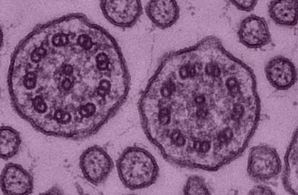
Cilia and flagella both have an internal structure built upon microtubules, but the flagellum is longer and is more often a single organelle. Inside both cilia and flagella is a microtubule-based cytoskeleton termed the axoneme, which provides scaffolding for various protein complexes. The transmission electron micrograph above left shows a cross-section through the axonemes of cilia with nine peripheral doublet microtubules and two central singlet microtubules (dynein arms stretch between the doublet microtubules – illustrations).
[] diagram comparing eu- and prokaryotic flagella : Microtubular arrangement down the length of a cilium or flagellum. Undulopodium. [] cross.gif (31K) ← description → axoneme.gif (90K) : 3D diagram - axoneme Џ 3D animation – inside flagellum [] image - detail of cilia : tem - structure cilium : diagram - mechanism of ciliary motility : Geometric Clutch Model Џ animation - cilia & flagella Џ animation - flagellum, lo-res [] col-sem cilia : tem - pulmonary cilia : tem - bacillus, flagella Џ Q-movie of nano-simulation of cap-directed flagellin assembly Џ
The motor protein dynein powers the sliding of the microtubules against one another — first on one side, then on the other. This dynein-powered sliding produces a whip-like motion employed to move fluid past or over the cell. During the course of evolution, cilia have been adapted to function as mechanoreceptors, chemoreceptors, and the outer segment of the rods in the vertebrate retina. Although called stereocilia, the hair-cell protrusions in the inner ear are actually modified villi. [] sem image - inner ear stereocilia []
The microtubular axoneme also provides binding sites for molecular motor proteins, such as kinesin II, which assist in the transport of proteins up and down the microtubules.
[] 3D diagram - axoneme Џ beautiful Flash 8 animation - Inner Life of the Cell and Interpretation: Inner Life of the Cell Џ
The microtubule organizing center, also called a centrosome, or a basal body, lies at the base of the cilium. The basal body, like the centriole, lacks the central pair and its peripheral microtubules are arranged in nine triplets rather than the doublets of the axoneme. [] diagram of flagellum : tem - basal bodies [] The basal body is created as the centriole (a microtubular structure essential to cell division) migrates to the surface. During animal cell division, centrosomes and centrioles replicate, generating two centrosomes, each with a pair of centrioles. The two centrosomes migrate to opposite sides of the nucleus and microtubules grow from the centrosome, becoming the spindle apparatus that separates replicated chromosomes into the two daughter cells.
The transition zone between axoneme and basal body serves as a docking station for intraflagellar transport and motor proteins. During intraflagellar transport (IFT) materials needed to build the cilia are carried to the ciliary tip and spent materials are carried down to the ciliary body. The IFT particle, which is made up of at least 17 polypeptide subunits, may also carry signals collected by various receptors embedded in the ciliary membrane.
▲ Top ▲
• A • adhesion • C • cell membranes • cellular signal transduction • centrioles • chemotaxis • chloroplast • cilia • communication • concentration gradients • cytokine receptors • cytoplasm • cytoskeleton • E • energy transducers • endoplasmic reticulum • endosomes • exosome • G • Golgi apparatus • GPCRs • H • hormones • I • ion channels • L • lysosome • M • meiosis • microtubules • mitosis • mitochondrion • N • Nitric Oxide • neurotransmission • neuronal interconnections • nuclear membrane • nuclear pore • P • pinocytosis • proteasome • pumps • R •receptor proteins • receptor-mediated endocytosis • S • second messengers • signaling gradients • signal transduction • spindle • structure • T • transport • two-component systems • V • vacuole • vesicle •
More: Cilia, Flagella, and Centrioles : Cilia and flagella : HHMI Bulletin September 2005: The Importance of Being Cilia : Google cilia : Virtual Cell Textbook - Cell Biology :