


microtubules
 Microtubules are cylindrical tubes 20-25 nm in diameter, which are composed of linear polymers (protofilaments) of the globular protein tubulin. (right - click to enlarge)
▼: active transport : cancer chemotherapy : centrioles : cilia, flagella, centrioles : conveyor belt : cycles assembly/disassembly : elongation : growth : growth factors : GTP : GTP and heterodimerization : heterodimers : MAPs : microtubule-associated proteins : mitotic spindle : nucleation : pathologies : polarity : protofilaments : scaffold : tau proteins : treadmilling : tubulin : XMAP 215 :▼
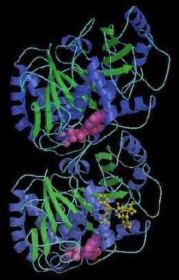
The tubulin molecules form heterodimers of α- and α-β-tubulin and linear rows of tubulin dimers form the protofilaments. Both α- and β-tubulins exist in several isotypic forms and can undergo several post-translational modifications. At least 14 tubulin isotypes that are expressed in a tissue specific manner have been identified in higher eukaryotes.
Microtubules act as scaffolding to maintain cell shape, and extend throughout the cytoplasm of eukaryotic cells. (Scaffold proteins that participate in signaling cascades are distinct from cytoskeletal scaffolding.) Microtubules are polar structures with two distinct ends, a fast growing "plus" end and a slow growing "minus" end. Microtubules are usually organized into a single array with their minus ends associated with a microtubule organizing center adjacent to the nucleus, and with their plus ends located toward the cell’s periphery near the plasma membrane. This arrangement establishes a defined polarity, which is utilized by the microtubule-associated motor proteins that move "cargo" to the minus or plus ends of cellular microtubules.
Because tubulin dimers constantly polymerize and depolymerize, microtubules can undergo rapid cycles of assembly and disassembly. The first stage of microtubules formation is the slow Mg2+ and GTP dependent "nucleation phase", in which α- and β-tubulins join end-to-end to form protofilaments with alternating α- and β-subunits. The second, elongation phase proceeds more rapidly.
GTP must be bound to both α- and β- subunits for tubulin heterodimerization and association of tubulins into microtubules. β-tubulin-bound-GTP is hydrolyzed to GDP during or immediately after polymerization, weakening the binding affinity of tubulin for adjacent molecules and favoring the depolymerization that contributes to the dynamic behavior of microtubules. Heterodimers can add onto or dissociate from either end of a microtubules, but there is greater tendency for addition to occur at the faster growing "plus" end where β-tubulin is exposed. Microtubules also undergo “treadmilling,” in which GTP-bound-tubulin molecules are continually lost from the minus end while being replaced by the addition of GTP-bound tubulin molecules to the plus end of the same microtubule.
During the microtubule formation, alternating elongation and shortening cycles provide dynamic instability that is critical for directing microtubules towards target sites, such as kinetochores, migrating membranes, and focal adhesions. Dynamic instability is a tightly regulated phenomenon, and is critical for the remodeling of the cytoskeleton during mitosis. Dynamic instability is characterized by four crucial variables:
a. rate of growth of microtubules,
b. rate of shortening of microtubules,
c. frequency of transition from the growth state to shortening, and
d. frequency of transition from shortening to growth.
The growth and shortening of microtubules is dependent upon the rate of tubulin addition relative to the rate of GTP hydrolysis. Tubulin-bound GTP is hydrolyzed to tubulin-GDP + Pi, yet tubulin-GTP can be added to the plus end almost simultaneously with its hydrolysis at the minus end. So, whenever GTP-bound tubulin molecules are added more rapidly than GTP is hydrolyzed, the microtubule will retain the GTP cap at its plus end and growth will continue. When the rate of polymerization decreases, the GTP that is bound to tubulin at the plus end is hydrolyzed to GDP and the GDP-bound tubulin dissociates, resulting in rapid depolymerization and shrinkage of microtubules.
Various proteins disassemble microtubules by increasing the rate of tubulin depolymerization. The inherent dynamic instability of microtubules can be modified by the interactions with microtubule-associated proteins (MAPs) and microtubule-regulatory proteins. The best-characterized MAPs are MAP-1, MAP-2, and tau proteins. MAPs can bind to microtubules, increasing their stability. The activity of MAPs is tightly regulated by their phosphorylation state. Altered phosphorylation state of MAPs has been positively linked to the pathogenesis of Alzheimer’s disease.
Growth factor signals activate protein kinases that catalyze phosphorylation of tubulin-binding domains of MAPs, causing them to detach from microtubules. XMAP215 is a highly conserved 215 kDa MAP, which plays an important role in controlling microtubular dynamics during the cell cycle. XMAP215 stabilizes the plus ends of microtubules, promoting elongation and preventing catastrophic shrinkage. At the onset of mitosis, higher phosphorylation of XMAP215 increases microtubular instability, causing disassembly. During the final phase of mitosis, protein phosphatase activity predominates as the microtubule array characteristic of interphase is re-established.
Because they play an essential role in the formation of the mitotic spindle during cell division, microtubules have been targetted for cancer chemotherapy. Anti-mitotic chemotherapeutic agents can selectively disrupt microtubular dynamics, either by targeting a specific tubulin isotype or targeting a particular stage of cell division. Such anti-mitotic agents exploit the difference in microtubular dynamics between normal cell populations and rapidly proliferating cancerous cells. For example, drugs such as colchicine and colcemid bind tubulin and inhibit microtubular polymerization, thus blocking mitosis. On the other hand, agents such as taxol stabilize microtubules, preventing cell division. [s] (diagram MTs, MAPs )
[] image_filamentous actin microtubules nuclei [] image_filamentous actin & microtubules [] image_microtubules nuclei endothelial tc [] image_filamentous actin microtubules nuclei fibroblast mouse [] image_tubulin microtubules Џ beautiful Flash 8 animation - Inner Life of the Cell, which shows interactions between adhesion-signaling molecules and the cytoskeleton, the scaffolding lattices and conveyor belt mechanisms, and assembly/disassembly of actin and tubulin, and Interpretation: Inner Life of the Cell Џ
In addition to their cytoskeletal roles, microtubules act as cellular conveyor belts, employing special attachment proteins to move chromosomes, granules, endosomal vesicles, and organelles such as mitochondria through the cytoplasm.
Microtubules may work alone, or be joined with other proteins to form more complex structures such as cilia and flagella and centrioles. Within cilia and flagella, microtubules are assembled in a 9 + 2 arrangement. animation - inside flagellum. The tubulin is coupled to dynein arms to enable locomotion (spermocytes, protozoa) or movement of liquid over the cell (important in embryologic differentiation). [] image_spermatozoa (mouse) [] image_sperm (Dv) [] diagram - mechanism of ciliary motility Џ animation - cilia & flagella
Arranged in orthogonal paired tubes of 9 fibers, microtubules form centrioles during cell division, or basal bodies at the root of cilia and flagella. animation - spinning centriole pair : tour centriole : zoom in on centriole. During mitosis microtubules form spindle fibres along which chromosomes assemble then separate.
Pathologies [s] associated with microtubules:
● dysfunction of stability
● dysfunction of microtubule-associated tau protein (taupathies)
● dysfunction of cellular cilia
● primary ciliary dyskinesia (DNAH5, DNAH7, DNAH11)
● anatomic lateralization defects (situs inversus)
● male infertility
● Kartagener syndrome - situs inversus with dextrocardia, bronchiectasis, chronic sinusitis, conductive deafness, immotile cilia and sperm
● pathology of microtubule-associated proteins (MAPs)
● pathology of tubulins and pathology of tubulin-specific chaperones
▲: active transport : cancer chemotherapy : centrioles סּ centrioles : cilia, flagella, centrioles סּ cilia : conveyor belt : cycles assembly/disassembly סּ cytoskeleton : elongation : growth : growth factors ~ growth factors : GTP : GTP and heterodimerization : heterodimers : MAPs microtubule-associated proteins : mitotic spindle סּ mitotic spindle : nucleation : polarity : protofilaments : scaffold סּ spindle : tau proteins : treadmilling : tubulin סּ vesicle : XMAP 215 :▲
Џ beautiful Flash 8 animation - inner life of the cell and description Џ animation - mitosis : animation ~ mitosis Џ link to animation - mitosis Џ kyrk animation _ mitosis [] image_mitosis microtubules kinetochores DNA [] image_mitosis [] image_aberrant division mammalian cell [] image_anaphase [] image_golgi apparatus DNA microtubules dividing cells [] image_mitotic spindle [] image - spindle Џ animation - mitosis animation - meiosis Џ kyrk animation _ meisosis : Google cytoskeleton : Google microtubule : Virtual Cell Textbook - Cell Biology :
• A • adhesion • C • cell membranes • cellular adhesion molecules • cellular signal transduction • centrioles • chemotaxis • chloroplast • cilia & flagella • communication • concentration gradients • cytokine receptors • cytoplasm • cytoskeleton • E • energy transducers • endoplasmic reticulum • endosomes • exosome • F • flagella & cilia • G • Golgi apparatus • GPCRs • H • hormones • I • ion channels • L • lysosome • M • meiosis • microtubules • mitosis • mitochondrion • N • Nitric Oxide • neurotransmission • neuronal interconnections • nuclear membrane • nuclear pore • P • pinocytosis • proteasome • pumps • R • receptor proteins • receptor-mediated endocytosis • S • second messengers • signaling gradients • signal transduction • spindle • structure • T • transport • two-component systems • V • vacuole • vesicle •